A synthetic peptide corresponding to a sequence at the N-terminus of human CD229/LY9, which shares 55.9% and 61.8% amino acid (aa) sequence identity with mouse and rat CD229/LY9, respectively.
Conjugation:
Unconjugated
Alternative Names:
T-lymphocyte surface antigen Ly-9, Cell surface molecule Ly-9, Lymphocyte antigen 9, SLAM family member 3, SLAMF3, Signaling lymphocytic activation molecule 3, CD229, LY9, CDABP0070
Anti-CD229/LY9 Antibody. Tested in Flow Cytometry, IHC, ICC applications. This antibody reacts with Human, Mouse, Rat.
Clonality:
Polyclonal
Concentration:
Adding 0.2 ml of distilled water will yield a concentration of 500 µg/ml.
Application Notes: Immunohistochemistry (Paraffin-embedded Section), 0.5-1µg/ml Immunohistochemistry (Frozen Section), 0.5-1µg/ml Immunocytochemistry, 0.5-1µg/ml Flow Cytometry (Fixed), 1-3µg/1x106 cells. Add 0.2ml of distilled water will yield a concentration of 500ug/ml
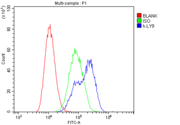
Flow Cytometry analysis of Daudi cells using anti-CD229 antibody. Overlay histogram showing Daudi cells (Blue line). To facilitate intracellular staining, cells were fixed with 4% paraformaldehyde and permeabilized with permeabilization buffer. The cells were blocked with 10% normal goat serum. And then incubated with rabbit anti-CD229 Antibody (1 µg/1x10 6 cells) for 30 min at 20C. DyLight488 conjugated goat anti-rabbit IgG (5-10 µg/1x10 6 cells) was used as secondary antibody for 30 minutes at 20C. Isotype control antibody (Green line) was rabbit IgG (1 µg/1x10 6) used under the same conditions. Unlabelled sample (Red line) was also used as a control.
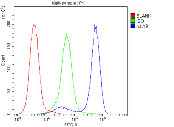
Flow Cytometry analysis of Jurkat cells using anti-CD229 antibody (
Flow Cytometry analysis of Jurkat cells using anti-CD229 antibody. Overlay histogram showing Jurkat cells (Blue line). To facilitate intracellular staining, cells were fixed with 4% paraformaldehyde and permeabilized with permeabilization buffer. The cells were blocked with 10% normal goat serum. And then incubated with rabbit anti-CD229 Antibody (1 µg/1x10 6 cells) for 30 min at 20C. DyLight488 conjugated goat anti-rabbit IgG (5-10 µg/1x10 6 cells) was used as secondary antibody for 30 minutes at 20C. Isotype control antibody (Green line) was rabbit IgG (1 µg/1x10 6) used under the same conditions. Unlabelled sample (Red line) was also used as a control.
Flow Cytometry analysis of U937 cells using anti-CD229 antibody. Overlay histogram showing U937 cells (Blue line). To facilitate intracellular staining, cells were fixed with 4% paraformaldehyde and permeabilized with permeabilization buffer. The cells were blocked with 10% normal goat serum. And then incubated with rabbit anti-CD229 Antibody (1 µg/1x10 6 cells) for 30 min at 20C. DyLight488 conjugated goat anti-rabbit IgG (5-10 µg/1x10 6 cells) was used as secondary antibody for 30 minutes at 20C. Isotype control antibody (Green line) was rabbit IgG (1 µg/1x10 6) used under the same conditions. Unlabelled sample (Red line) was also used as a control.
IHC analysis of CD229 using anti-CD229 antibody. CD229 was detected in paraffin-embedded section of mouse spleen tissues. Heat mediated antigen retrieval was performed in citrate buffer (pH6, epitope retrieval solution) for 20 mins. The tissue section was blocked with 10% goat serum. The tissue section was then incubated with 1 µg/ml rabbit anti-CD229 Antibody overnight at 4C. Biotinylated goat anti-rabbit IgG was used as secondary antibody and incubated for 30 minutes at 37C. The tissue section was developed using Strepavidin-Biotin-Complex (SABC) with DAB as the chromogen.
IHC analysis of CD229 using anti-CD229 antibody. CD229 was detected in paraffin-embedded section of mouse spleen tissues. Heat mediated antigen retrieval was performed in citrate buffer (pH6, epitope retrieval solution) for 20 mins. The tissue section was blocked with 10% goat serum. The tissue section was then incubated with 1 µg/ml rabbit anti-CD229 Antibody overnight at 4C. Biotinylated goat anti-rabbit IgG was used as secondary antibody and incubated for 30 minutes at 37C. The tissue section was developed using Strepavidin-Biotin-Complex (SABC) with DAB as the chromogen.
IHC analysis of CD229 using anti-CD229 antibody. CD229 was detected in paraffin-embedded section of rat spleen tissues. Heat mediated antigen retrieval was performed in citrate buffer (pH6, epitope retrieval solution) for 20 mins. The tissue section was blocked with 10% goat serum. The tissue section was then incubated with 1 µg/ml rabbit anti-CD229 Antibody overnight at 4C. Biotinylated goat anti-rabbit IgG was used as secondary antibody and incubated for 30 minutes at 37C. The tissue section was developed using Strepavidin-Biotin-Complex (SABC) with DAB as the chromogen.
IHC analysis of CD229 using anti-CD229 antibody. CD229 was detected in paraffin-embedded section of rat spleen tissues. Heat mediated antigen retrieval was performed in citrate buffer (pH6, epitope retrieval solution) for 20 mins. The tissue section was
* VAT and and shipping costs not included. Errors and price changes excepted