zeta chain of T cell receptor associated protein kinase 70
Application Notes:
Application Notes: Western blot, 0.1-0.5µg/ml Immunohistochemistry (Paraffin-embedded Section), 0.5-1µg/ml. Add 0.2ml of distilled water will yield a concentration of 500µg/ml
IHC analysis of Collagen III/COL3A1 using anti-Collagen III/COL3A1 antibody. Collagen III/COL3A1 was detected in paraffin-embedded section of human intestinal cancer tissue. Heat mediated antigen retrieval was performed in EDTA buffer (pH8.0, epitope retrieval solution). The tissue section was blocked with 10% goat serum. The tissue section was then incubated with 1 µg/ml mouse anti-Collagen III/COL3A1 Antibody overnight at 4C. Biotinylated goat anti-mouse IgG was used as secondary antibody and incubated for 30 minutes at 37C. The tissue section was developed using Strepavidin-Biotin-Complex (SABC) with DAB as the chromogen.
IHC analysis of Collagen III/COL3A1 using anti-Collagen III/COL3A1 antibody. Collagen III/COL3
IHC analysis of Collagen III/COL3A1 using anti-Collagen III/COL3A1 antibody. Collagen III/COL3A1 was detected in paraffin-embedded section of human intestinal cancer tissue. Heat mediated antigen retrieval was performed in EDTA buffer (pH8.0, epitope retrieval solution). The tissue section was blocked with 10% goat serum. The tissue section was then incubated with 1 µg/ml mouse anti-Collagen III/COL3A1 Antibody overnight at 4C. Biotinylated goat anti-mouse IgG was used as secondary antibody and incubated for 30 minutes at 37C. The tissue section was developed using Strepavidin-Biotin-Complex (SABC) with DAB as the chromogen.
IHC analysis of Collagen III/COL3A1 using anti-Collagen III/COL3A1 antibody. Collagen III/COL3A1 was detected in paraffin-embedded section of human lung cancerr tissue. Heat mediated antigen retrieval was performed in EDTA buffer (pH8.0, epitope retrieval solution). The tissue section was blocked with 10% goat serum. The tissue section was then incubated with 1 µg/ml mouse anti-Collagen III/COL3A1 Antibody overnight at 4C. Biotinylated goat anti-mouse IgG was used as secondary antibody and incubated for 30 minutes at 37C. The tissue section was developed using Strepavidin-Biotin-Complex (SABC) with DAB as the chromogen.
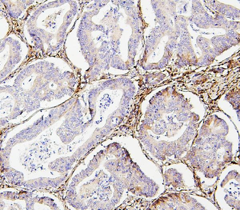
IHC analysis of Collagen III/COL3A1 using anti-Collagen III/COL3A1 antibody. Collagen III/COL3A1 was detected in paraffin-embedded section of human mammary cancer tissue. Heat mediated antigen retrieval was performed in EDTA buffer (pH8.0, epitope retrieval solution). The tissue section was blocked with 10% goat serum. The tissue section was then incubated with 1 µg/ml mouse anti-Collagen III/COL3A1 Antibody overnight at 4C. Biotinylated goat anti-mouse IgG was used as secondary antibody and incubated for 30 minutes at 37C. The tissue section was developed using Strepavidin-Biotin-Complex (SABC) with DAB as the chromogen.
IHC analysis of Collagen III/COL3A1 using anti-Collagen III/COL3A1 antibody. Collagen III/COL3A1 was detected in paraffin-embedded section of mouse intestine tissue. Heat mediated antigen retrieval was performed in EDTA buffer (pH8.0, epitope retrieval solution). The tissue section was blocked with 10% goat serum. The tissue section was then incubated with 1 µg/ml mouse anti-Collagen III/COL3A1 Antibody overnight at 4C. Biotinylated goat anti-mouse IgG was used as secondary antibody and incubated for 30 minutes at 37C. The tissue section was developed using Strepavidin-Biotin-Complex (SABC) with DAB as the chromogen.
IHC analysis of Collagen III/COL3A1 using anti-Collagen III/COL3A1 antibody. Collagen III/COL3A1 was detected in paraffin-embedded section of rat intestine tissue. Heat mediated antigen retrieval was performed in EDTA buffer (pH8.0, epitope retrieval solution). The tissue section was blocked with 10% goat serum. The tissue section was then incubated with 1 µg/ml mouse anti-Collagen III/COL3A1 Antibody overnight at 4C. Biotinylated goat anti-mouse IgG was used as secondary antibody and incubated for 30 minutes at 37C. The tissue section was developed using Strepavidin-Biotin-Complex (SABC) with DAB as the chromogen.
Western blot analysis of Collagen III/COL3A1 using anti-Collagen III/COL3A1 antibody. Electrophoresis was performed on a 5-20% SDS-PAGE gel at 70V (Stacking gel) / 90V (Resolving gel) for 2-3 hours. The sample well
* VAT and and shipping costs not included. Errors and price changes excepted