136.36mM Ethanolamine, 133.23 mM Chlorides, 9.55mM Phosphates, 9.55mM Sodium Bicarbonate.
Target:
ATG7
Application Dilute:
WB (1:1000), ICC/IF (1:100), IHC (1:50)
Application Notes:
Application Notes: A 1:1000 dilution of SPC-610 was sufficient for detection of ATG7 in 15 µg of human HeLa cell lysates by ECL immunoblot analysis using goat anti-rabbit IgG:HRP as the secondary antibody
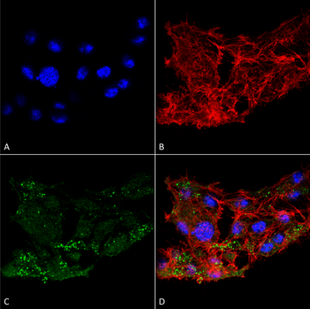
Immunocytochemistry/Immunofluorescence analysis using Rabbit Anti-ATG7 Polyclonal Antibody. Tissue: Colon carcinoma cell line (RKO). Species: Human. Fixation: 4% Formaldehyde for 15 min at RT. Primary Antibody: Rabbit Anti-ATG7 Polyclonal Antibody at 1:100 for 60 min at RT. Secondary Antibody: Goat Anti-Rabbit ATTO 488 at 1:100 for 60 min at RT. Counterstain: Phalloidin Texas Red F-Actin stain, DAPI (blue) nuclear stain at 1:1000, 1:5000 for 60 min at RT, 5 min at RT. Localization: Cytoplasm, Preautophagosomal Structure, Organelle membrane. Magnification: 60X. (A) DAPI nuclear stain. (B) Phalloidin Texas Red F-Actin stain. (C) ATG7 Antibody. (D) Composite.
Immunohistochemistry analysis using Rabbit Anti-ATG10 Polyclonal Antibody. Tissue: Brain. Species: Human. Fixation: Formalin Fixed Paraffin-Embedded. Primary Antibody: Rabbit Anti-ATG10 Polyclonal Antibody at 1:50 for 31 min at RT. Counterstain: Hematoxylin. Magnification: 10X.
Western blot analysis of Rat brain cell lysates showing detection of ~77.9 kDa ATG7 protein using Rabbit Anti-ATG7 Polyclonal Antibody. Lane 1: Molecular Weight Ladder (MW). Lane 2: Rat brain cell lysates. Load: 20 µg. Block: 2% BSA and 2% Skim Milk in 1X TBST. Primary Antibody: Rabbit Anti-ATG7 Polyclonal Antibody at 1:1000 for 16 hours at 4C. Secondary Antibody: Goat Anti-Rabbit IgG: HRP at 1:2000 for 60 min at RT. Color Development: ECL solution for 6 min at RT. Predicted/Observed Size: ~77.9 kDa.
Western blot analysis of Human Cervical cancer cell line (HeLa) lysate showing detection of ~77.9 kDa ATG7 protein using Rabbit Anti-ATG7 Polyclonal Antibody. Lane 1: Molecular Weight Ladder (MW). Lane 2: Human Cervical cancer cell line (HeLa) lysate. Load: 15 µg. Block: 5% Skim Milk in 1X TBST. Primary Antibody: Rabbit Anti-ATG7 Polyclonal Antibody at 1:1000 for 60 min at RT. Secondary Antibody: Goat Anti-Rabbit IgG: HRP at 1:2000 for 60 min at RT. Color Development: ECL solution for 6 min at RT. Predicted/Observed Size: ~77.9 kDa.
* VAT and and shipping costs not included. Errors and price changes excepted