Liquid. Purified antibody supplied in 1x PBS buffer with 0.09% (w/v) sodium azide and 2% sucrose.
Form:
Liquid. Purified antibody supplied in 1x PBS buffer with 0.09% (w/v) sodium azide and 2% sucrose.
Sequence:
Synthetic peptide located within the following region: RRQLTKPRPVILDPADPTGNLGGGDPKGWRQLAQEAEAWLNYPCFKNWDG
Target:
OAS1
Application Notes:
Application Notes: Application Info: IHC - Paraffin ~~ 5 ug/ml Western blot ~~1 ug/ml
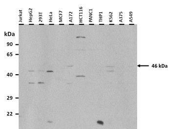
25 ug of the indicated Human whole cell or tissue extracts was loaded onto a 12% SDS-PAGE gel. 3 ug/ml of the antibody was used in this experiment. Peptide is present in isoforms of 47, 46, 42 and 41 kDa.
Sample Tissue: Human Hela Whole Cell, Antibody dilution: 3 ug/ml.
Sample Tissue: Human Hela, Antibody dilution: 1.0 ug/ml.
Sample Tissue: Human MCF7 Whole Cell, Antibody dilution: 3 ug/ml.
Positive control (+): HepG2 (HG), Negative control (-): 293T (2T), Antibody concentration: 1 ug/ml.
Immunohistochemistry of formalin-fixed, paraffin-embedded human Small intestine tissue. Antibody concentration 5 ug/ml.
Surface Plasmon Resonance Kinetic Characterization of Polyclonal Antibody Affinity. Purified polyclonal antibodies were immobilized on a Protein A/G coated Carterra LSA sensor chip (PAGH200M) at concentrations of 5, and 50 ug/ml in duplicate. Antibodies on the surface were exposed to interaction with peptides sequentially via microfluidic controlled flow at 333nM peptide concentration for kinetic characterization of the binders for affinity and specificity, followed by curve fitting using the Kinetics software. Kd determinations for both concentrations were averaged and results and standard deviation are shown.