
Phospho-CDK1 (Thr14) Rabbit Polyclonal Antibody, Unconjugated
Catalog Number:
BYT-ORB5822
- Images (6)
| Article Name: | Phospho-CDK1 (Thr14) Rabbit Polyclonal Antibody, Unconjugated |
| Biozol Catalog Number: | BYT-ORB5822 |
| Supplier Catalog Number: | orb5822 |
| Alternative Catalog Number: | BYT-ORB5822-100, BYT-ORB5822-200, BYT-ORB5822-50 |
| Manufacturer: | Biorbyt |
| Host: | Rabbit |
| Category: | Antikörper |
| Application: | FC, ICC, IF, IHC-Fr, IHC-P, WB |
| Species Reactivity: | Human, Mouse, Rat |
| Immunogen: | KLH conjugated Synthesised phosphopeptide derived from human cdc2 around the phosphorylation site of Thr14 EG(p-T)YG |
| Conjugation: | Unconjugated |
| Alternative Names: | CDK1 (p-T14), p-CDK1, phospho-CDK1, CDC2, CDC28A, P34CDC2, Cdc2a, p34, CDK1_CHICK, CDK1, Cell division control protein 2 homolog, Cell division protein kinase 1, p34 protein kinase, 2.7.11.22, 2.7.11.23, CDK1_HUMAN, CDKN1, CDK1_MOUSE, CDK1_RAT, |
| Phospho-CDK1 (Thr14) Rabbit Polyclonal Antibody |
| Clonality: | Polyclonal |
| Concentration: | 1mg/ml |
| Molecular Weight: | 34 kDa |
| UniProt: | P06493 |
| Buffer: | 0.01M TBS (pH7.4) with 1% rAlbumin, 0.02% Proclin300 and 50% Glycerol. |
| Form: | Liquid |
| Target: | CDK1 |
| Application Dilute: | WB=1:500-2000, IHC-P=1:100-500, IHC-F=1:100-500, ICC/IF=1:100-500, IF=1:100-500, Flow-Cyt=1ug/Test |
| Application Notes: | Modification: Phosphorylated |
|
|
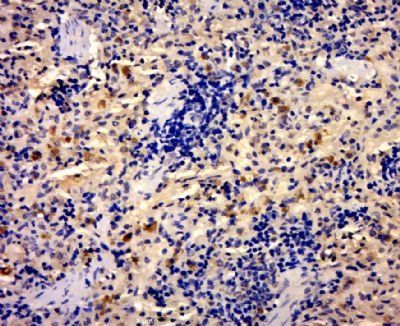
Immunohistochemical staining of rat spleen tissue using cdc2 (phospho-Thr14) antibody. |
|
|
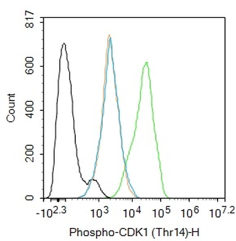
Blank control (black line): Hela. Primary Antibody (green line): Rabbit Anti-Phospho-CDK1 (Thr14) antibody (orb5822), dilution: 1 ug/Test, Secondary Antibody (white blue line): Goat anti-rabbit IgG-AF488, dilution: 0.5 ug/Test. Isotype control (orange line): Normal Rabbit IgG, Protocol, The cells were fixed with 4% PFA (10 min at room temperature) and then permeabilized with 90% ice-cold methanol for 20 min at -20C, The cells were then incubated in 5% BSA to block non-specific protein-protein interactions for 30 min at room temperature. Cells stained with Primary Antibody for 30 min at room temperature. The secondary antibody used for 40 min at room temperature. Acquisition of 20000 events was performed. |
|
|
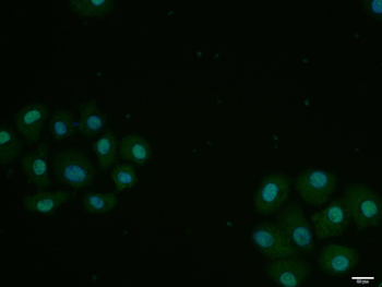
HepG2 cell, 4% Paraformaldehyde-fixed, Triton X-100 at room temperature for 20 min, Blocking buffer (normal goat serum) at 37C for 20 min, Antibody incubation with (Phospho-CDK1 (Thr14)) polyclonal Antibody, Unconjugated (orb5822) 1:100, 90 minutes at 37C, followed by a conjugated Goat Anti-Rabbit IgG antibody at 37C for 90 minutes, DAPI (blue) was used to stain the cell nuclei. |
|
|
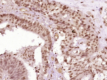
Paraformaldehyde-fixed, paraffin embedded (Human colon cancer), Antigen retrieval by boiling in sodium citrate buffer (pH6.0) for 15 min, Block endogenous peroxidase by 3% hydrogen peroxide for 20 minutes, Blocking buffer (normal goat serum) at 37C for 30 min, Antibody incubation with (Phospho-CDK1 (Thr14)) Polyclonal Antibody, Unconjugated (orb5822) at 1:400 overnight at 4C, followed by operating according to SP Kit (Rabbit) instructions and DAB staining. |
|
|
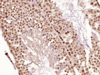
Paraformaldehyde-fixed, paraffin embedded (Mouse testis), Antigen retrieval by boiling in sodium citrate buffer (pH6.0) for 15 min, Block endogenous peroxidase by 3% hydrogen peroxide for 20 minutes, Blocking buffer (normal goat serum) at 37C for 30 min, Antibody incubation with (Phospho-CDK1 (Thr14)) Polyclonal Antibody, Unconjugated (orb5822) at 1:400 overnight at 4C, followed by operating according to SP Kit (Rabbit) instructions and DAB staining. |
|
|
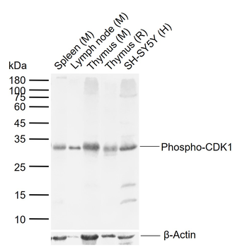
Sample: Lane 1: Mouse Spleen tissue lysates, Lane 2: Mouse Lymph node tissue lysates, Lane 3: Mouse Thymus tissue lysates, Lane 4: Rat Thymus tissue lysates, Lane 5: Human SH-SY5Y cell lysates, Primary: Anti-Phospho-CDK1 (Thr14) (orb5822) at 1/1000 dilution, Secondary: IRDye800CW Goat Anti-Rabbit IgG at 1/20000 dilution, Predicted band size: 34 kDa, Observed band size: 32 kDa. |
Product Guarantee and Expert Support
