136.36mM Ethanolamine, 133.23 mM Chlorides, 9.55mM Phosphates, 9.55mM Sodium Bicarbonate
Target:
Thyroid Hormone Receptor
Application Dilute:
WB (1:500), IHC (1:100)
Application Notes:
Application Notes: A 1:500 dilution of SMC-561 was sufficient for detection of Pan-Thyroid hormone receptor in 10 ug of Hep G2 Human Hepatoblastoma Cell lysate by ECL immunoblot analysis using goat anti-mouse IgG:HRP as the secondary antibody
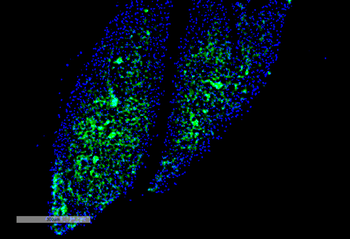
Immunohistochemistry analysis using Mouse Anti-Thyroid Hormone Receptor Monoclonal Antibody, Clone H43. Tissue: Thyroid. Species: Mouse. Primary Antibody: Mouse Anti-Thyroid Hormone Receptor Monoclonal Antibody at 1:100 for Overnight at 4C, then 30 min at 37C. Secondary Antibody: Goat Anti-Mouse IgG (H+L): FITC for 45 min at 37C. Counterstain: DAPI for 3 min at RT. Magnification: 7.5X.
Immunohistochemistry analysis using Mouse Anti-Thyroid Hormone Receptor Monoclonal Antibody, Clone H43. Tissue: Thyroid Cancer. Species: Human. Primary Antibody: Mouse Anti-Thyroid Hormone Receptor Monoclonal Antibody at 1:100 for Overnight at 4C, then 30 min at 37C. Secondary Antibody: Goat Anti-Mouse IgG (H+L): FITC for 45 min at 37C. Counterstain: DAPI for 3 min at RT. Magnification: 4X.
Western Blot analysis of Human Hep G2 Hepatoblastoma Cell lysate showing detection of Thyroid Hormone Receptor protein using Mouse Anti-Thyroid Hormone Receptor Monoclonal Antibody, Clone H43. Load: 10 ug. Primary Antibody: Mouse Anti-Thyroid Hormone Receptor Monoclonal Antibody at 1:500 for 2 hours at RT with shaking. Secondary Antibody: Goat anti-mouse IgG:HRP at 1:4000 for 1 hour at RT with shaking. Color Development: Chemiluminescent for HRP (Moss) for 5 min in RT. Other Band (s): Higher molceular weight bands could be due to PTMs.
* VAT and and shipping costs not included. Errors and price changes excepted