
Tau (pSer202/ pThr205) Antibody (Biotin), Clone: [AH36], Rabbit, Monoclonal
Catalog Number:
BYT-ORB612744
- Images (5)


| Article Name: | Tau (pSer202/ pThr205) Antibody (Biotin), Clone: [AH36], Rabbit, Monoclonal |
| Biozol Catalog Number: | BYT-ORB612744 |
| Supplier Catalog Number: | orb612744 |
| Alternative Catalog Number: | BYT-ORB612744-100 |
| Manufacturer: | Biorbyt |
| Host: | Rabbit |
| Category: | Antikörper |
| Application: | DOT, ELISA, ICC, IF, IHC, WB |
| Species Reactivity: | Human, Mouse |
| Immunogen: | Synthetic peptide of Human Phospho Tau (Ser202/Thr205) |
| Conjugation: | Biotin |
| Alternative Names: | Tau, TAU, TAU_HUMAN, MAPT, MAPTL, Microtubule-associated protein tau, Microtubule associated protein tau, Microtubule associated protein tau isoform 4, Neurofibrillary tangle protein, Paired helical filament tau, Paired helical filament-tau, PHF tau, PHF-tau, DDPAC, FTDP 17, G protein beta1/gamma2 subunit interacting factor 1, MSTD, Mtapt, MTBT1, MTBT2, PPND, PPP1R103, Protein phosphatase 1 regulatory subunit 103, RNPTAU, AI413597, AW045860, FLJ31424, MGC134287, MGC138549, MGC156665 |
| Rabbit monoclonal antibody against Tau conjugated to Biotin |
| Clonality: | Monoclonal |
| Concentration: | 1 mg/ml |
| Clone Designation: | [AH36] |
| Molecular Weight: | 79 kDa |
| UniProt: | P10636 |
| Buffer: | 136.36mM Ethanolamine, 133.23 mM Chlorides, 9.55mM Phosphates, 9.55mM Sodium Bicarbonate |
| Target: | Tau (pSer202/ pThr205) |
| Application Dilute: | WB (1:500), ICC/IF (1:500), IHC (1:500) |
| Application Notes: | Application Notes: A 1:500 dilution of SMC-601 was sufficient for detection of Tau in 10 µg Baculovirus by dot blot analysis using Goat Anti-Rabbit IgG:HRP as the secondary antibody |
|
|
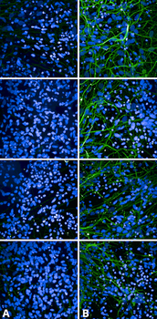
Immunocytochemistry/Immunofluorescence analysis using Rabbit Anti-Tau Monoclonal Antibody, Clone AH36. Tissue: iPSC-derived cortical excitatory neurons. Species: Human. Primary Antibody: Rabbit Anti-Tau Monoclonal Antibody at 1:500 for Overnight. Secondary Antibody: Donkey anti-rabbit: Alexa Fluor 488 at 1:1000. Counterstain: DAPI. A) iPSC-derived neurons from non-demented control (NDC). B) iPSC-derived neurons from subject with P301L MAPT mutation. Images acquired using an automated Opera Phoenix system. Each field of view is a max projection from 10 planes of 1 µm stacks. |
|
|
Immunohistochemistry analysis using Rabbit Anti-Tau Monoclonal Antibody, Clone AH36. Tissue: Brain slice. Species: Mouse. Primary Antibody: Rabbit Anti-Tau Monoclonal Antibody at 1:500 for Overnight at 4C. Secondary Antibody: Anti-Rabbit IgG: AlexaFluor 488. Counterstain: DAPI at 1:1000 for 5 min. (A) Pons of Non-Tg mouse. (B) Pons of P301SxUBQLN2 Tg mouse. (C) Prefrontal cortex of Non-Tg mouse. (D) Prefrontal cortex of P301SxUBQLN2 Tg mouse. IHC Protocol: 1. Post-fix brains in 4% PFA for 24 hours and put through a 10-30% sucrose gradient. 2. Section by cryostat at 10 uM thickness. 3. Fix in MeOH 15 min. 4. 3x10 min wash in PBS 1X. 5. Heat via microwave in 10mM Citrate Buffer, pH 6 for 4 min at power level 20. 6. Cool in solution for 20 min. 7. Wash 2x5 min in PBS. 8. Permeabilize in 0.5% Triton-X 100 in PBS 10 min. 9. Wash in PBS 10 min. 10. Block for 1 hour in 5% goat serum. 11. Incubate primary Ab (at 1:500) in blocking solution overnight at 4C. 12. Wash 3x10 min in PBS. 13. Incubate in secondary Ab Rb IgG Alexa-fluor 488. 14. Wash 3x10 min in PBS. 15. Incubate in DAPI 1:1000 for 5 min. 16. Wash 3x5 min. 17. Coverslip with Prolong-Gold. |
|
|
Immunohistochemistry analysis using Rabbit Anti-Tau Monoclonal Antibody, Clone AH36. Tissue: Brain slice. Species: Mouse. Primary Antibody: Rabbit Anti-Tau Monoclonal Antibody at 1:500 for Overnight at 4C. Secondary Antibody: Anti-Rabbit IgG: AlexaFluor 488. Counterstain: DAPI at 1:1000 for 5 min. CA3 Region of P301SxUBQLN2 Tg mouse. IHC Protocol: 1. Post-fix brains in 4% PFA for 24 hours and put through a 10-30% sucrose gradient. 2. Section by cryostat at 10 uM thickness. 3. Fix in MeOH 15 min. 4. 3x10 min wash in PBS 1X. 5. Heat via microwave in 10mM Citrate Buffer, pH 6 for 4 min at power level 20. 6. Cool in solution for 20 min. 7. Wash 2x5 min in PBS. 8. Permeabilize in 0.5% Triton-X 100 in PBS 10 min. 9. Wash in PBS 10 min. 10. Block for 1 hour in 5% goat serum. 11. Incubate primary Ab (at 1:500) in blocking solution overnight at 4C. 12. Wash 3x10 min in PBS. 13. Incubate in secondary Ab Rb IgG Alexa-fluor 488. 14. Wash 3x10 min in PBS. 15. Incubate in DAPI 1:1000 for 5 min. 16. Wash 3x5 min. 17. Coverslip with Prolong-Gold. |
|
|
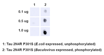
Dot Blot analysis using Rabbit Anti-Tau Monoclonal Antibody, Clone AH36. Species: E. Coli, Baculovirus. Primary Antibody: Rabbit Anti-Tau Monoclonal Antibody at 1:500. Secondary Antibody: Goat anti-rabbit IgG:HRP. |
|
|
Western Blot analysis of Human iPSC-derived cortical neurons showing detection of Tau protein using Rabbit Anti-Tau Monoclonal Antibody, Clone AH36. Lane 1: MW ladder. Lane 2: Control (non-disease) line. Lane 2: Ex10+16 tau mutant sample. Lane 3: P301L tau mutant sample. Load: 50ug. Primary Antibody: Rabbit Anti-Tau Monoclonal Antibody at 1:500 for Overnight. Total tau was detected using mouse anti-tau antibody (clone HT7). The bar graph on the right shows quantification of pSer202/pThr205 compared to total tau in each sample. |
Product Guarantee and Expert Support
