136.36mM Ethanolamine, 133.23 mM Chlorides, 9.55mM Phosphates, 9.55mM Sodium Bicarbonate
Target:
VPS35
Application Dilute:
WB (1:1000), ICC/IF (1:200), IP (1:200), IHC (1:100)
Application Notes:
Application Notes: A 1:1000 dilution of SMC-604 was sufficient for detection of VPS35 in 10 µg of SH-SY5Y by ECL immunoblot analysis using Goat Anti-Mouse IgG:HRP as the secondary antibody
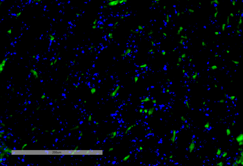
Immunohistochemistry analysis using Mouse Anti-VPS35 Monoclonal Antibody, Clone 8A3. Tissue: Kidney. Species: Mouse. Primary Antibody: Mouse Anti-VPS35 Monoclonal Antibody at 1:100 for Overnight at 4C, then 30 min at 37C. Secondary Antibody: Goat Anti-Mouse IgG (H+L): FITC for 45 min at 37C. Counterstain: DAPI for 3 min at RT. Magnification: 20X.
Immunocytochemistry/Immunofluorescence analysis using Mouse Anti-VPS35 Monoclonal Antibody, Clone 8A3. Tissue: A549 cells. Species: Human. Primary Antibody: Mouse Anti-VPS35 Monoclonal Antibody at 1:5 (tissue culture supernatant). Secondary Antibody: Donkey anti-mouse: Alexa Fluor 594 at 1:4000 in 0.2% BSA PBS. Counterstain: DAPI. Localization: Vesicles. A) VPS35 KO A549 cells B) WT A549 cells.
Immunocytochemistry/Immunofluorescence analysis using Mouse Anti-VPS35 Monoclonal Antibody, Clone 8A3. Tissue: A549 WT, VPS35 KO cells. Species: Human. Primary Antibody: Mouse Anti-VPS35 Monoclonal Antibody. Secondary Antibody: Donkey Anti-Mouse AlexaFluor 594. Clone can detect VPS35 at 1/2000 concentration.
Western Blot analysis of Human, Mouse A549, MEF showing detection of VPS35 protein using Mouse Anti-VPS35 Monoclonal Antibody, Clone 8A3. Lane 1: Molecular Weight Ladder. Lane 2: VPS35 KO A549 cells. Lane 3: mouse embryonic fibroblast cells. Load: 8 µg each A549 and MEF. Primary Antibody: Mouse Anti-VPS35 Monoclonal Antibody at 1:5 (tissue culture supernatant). Secondary Antibody: Donkey anti-mouse IRDye 800CW at 1:25000 in TBS-T.
Western Blot analysis of Human SH-SY5Y showing detection of VPS35 protein using Mouse Anti-VPS35 Monoclonal Antibody, Clone 8A3. Lane 1: Molecular Weight Ladder. Lane 2: SH-SY5Y (10 ug). Load: 10 µg. Block: 5% Skim Milk powder in TBST. Primary Antibody: Mouse Anti-VPS35 Monoclonal Antibody at 1:1000 for 2 hours at RT with shaking. Secondary Antibody: Goat anti-mouse IgG:HRP at 1:4000 for 1 hour at RT with shaking. Color Development: Chemiluminescent for HRP (Moss) for 5 min in RT.
Immunoprecipitation analysis using Mouse Anti-VPS35 Monoclonal Antibody, Clone 8A3. Tissue: A549 cells. Species: Human. Primary Antibody: Mouse Anti-VPS35 Monoclonal Antibody. Three amounts of (3, 1 and 0.3 ug) were non-covalently coupled to 10uL of A/G sepharose beads for 1 hour at 4C and next incubated with 250ug of A549 lysate for 2 hours at 4C.
Immunoprecipitation analysis using Mouse Anti-VPS35 Monoclonal Antibody, Clone 8A3. Tissue: embryonic fibroblast. Species: Mouse. Primary Antibody: Mouse Anti-VPS35 Monoclonal Antibody. Three amounts of (3, 1 and 0.3 ug) were non-covalently coupled to 10uL of A/G sepharose beads for 1 hour at 4C and next incubated with 250ug of MEF lysate for 2 hours at 4C.
Immunoprecipitation analysis using Mouse Anti-VPS35 Monoclonal Antibody, Clone 8A3. Tissue: A549 cells. Species: Human. Primary Antibody: Mouse Anti-VPS35 Monoclonal Antibody. 500 µL cell culture supernatants were incubated with 10 µL of Protein A/G resin beads for 1 hour at 4C.
* VAT and and shipping costs not included. Errors and price changes excepted