Each vial contains 4mg Trehalose, 0.9mg NaCl and 0.2mg Na2HPO4.
Form:
Lyophilized
Target:
ATP-dependent RNA helicase DHX15
Application Dilute:
Western blot, 0.25-0.5µg/ml, Human, Mouse, Rat Immunohistochemistry (Paraffin-embedded Section), 2-5µg/ml, Human Immunocytochemistry/Immunofluorescence, 5µg/ml, Human Flow Cytometry(Fixed), 1-3µg/1x10 6 cells, Human
Flow Cytometry analysis of Hela cells using anti-DHX15/prp43 antibody. Overlay histogram showing Hela cells (Blue line). To facilitate intracellular staining, cells were fixed with 4% paraformaldehyde and permeabilized with permeabilization buffer. The cells were blocked with 10% normal goat serum. And then incubated with rabbit anti-DHX15/prp43 Antibody (1 µg/1x10 6 cells) for 30 min at 20C. DyLight488 conjugated goat anti-rabbit IgG (5-10 µg/1x10 6 cells) was used as secondary antibody for 30 minutes at 20C. Isotype control antibody (Green line) was rabbit IgG (1 µg/1x10 6) used under the same conditions. Unlabelled sample (Red line) was also used as a control.
IF analysis of DHX15/prp43 using anti-DHX15/prp43 antibody.
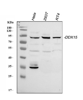
WB analysis using anti-DHX15/prp43 antibody.Lane 1:HeLa cell, Lane 2:293T cell, Lane 3:RT4 cell.
IF analysis of DHX15/prp43 using anti-DHX15/prp43 antibody and anti-Beta Tubulin antibody. DHX15/prp43 was detected in immunocytochemical section of U2OS cell. Enzyme antigen retrieval was performed using IHC enzyme antigen retrieval reagent for 15 mins. The cells were blocked with 10% goat serum. And then incubated with 5 µg/mL rabbit anti-DHX15/prp43 Antibody and mouse anti-Beta Tubulin antibody overnight at 4C. DyLight488 Conjugated Goat Anti-Rabbit IgG and Cy3 Conjugated Goat Anti-Mouse IgG were used as secondary antibody at 1:500 dilution and incubated for 30 minutes at 37C. Visualize using a fluorescence microscope and filter sets appropriate for the label used.
IHC analysis of DHX15/prp43 using anti-DHX15/prp43 antibody. DHX15/prp43 was detected in a paraffin-embedded section of human colorectal adenocarcinoma tissue. Heat mediated antigen retrieval was performed in EDTA buffer (pH8.0, epitope retrieval solution). The tissue section was blocked with 10% goat serum. The tissue section was then incubated with 2 µg/ml rabbit anti-DHX15/prp43 Antibody overnight at 4C. Peroxidase Conjugated Goat Anti-rabbit IgG was used as secondary antibody and incubated for 30 minutes at 37C. The tissue section was developed using HRP Conjugated Rabbit IgG Super Vision Assay Kit with DAB as the chromogen.
IHC analysis of DHX15/prp43 using anti-DHX15/prp43 antibody. DHX15/prp43 was detected in a paraffin-embedded section of human liver cancer tissue. Heat mediated antigen retrieval was performed in EDTA buffer (pH8.0, epitope retrieval solution). The tissue section was blocked with 10% goat serum. The tissue section was then incubated with 2 µg/ml rabbit anti-DHX15/prp43 Antibody overnight at 4C. Peroxidase Conjugated Goat Anti-rabbit IgG was used as secondary antibody and incubated for 30 minutes at 37C. The tissue section was developed using HRP Conjugated Rabbit IgG Super Vision Assay Kit with DAB as the chromogen.
IHC analysis of DHX15/prp43 using anti-DHX15/prp43 antibody. DHX15/prp43 was detected in a paraffin-embedded section of human prostate cancer tissue. Heat mediated antigen retrieval was performed in EDTA buffer (pH8.0, epitope retrieval solution). The tissue section was blocked with 10% goat serum. The tissue section was then incubated with 2 µg/ml rabbit anti-DHX15/prp43 Antibody overnight at 4C. Peroxidase Conjugated Goat Anti-rabbit IgG was used as secondary antibody and incubated for 30 minutes at 37C. The tissue section was developed using HRP Conjugated Rabbit IgG Super Vision Assay Kit with DAB as the chromogen.
IHC analysis of DHX15/prp43 using anti-DHX15/prp43 antibody. DHX15/prp43 was detected in a paraffin-embedded section of human testicular germ cell tumor tissue. Heat mediated antigen retrieval was performed in EDTA buffer (pH8.0, epitope retrieval solution). The tissue section was blocked with 10% goat serum. The tissue section was then incubated with 2 µg/ml rabbit anti-DHX15/prp43 Antibody overnight at 4C. Peroxidase Conjugated Goat Anti-rabbit IgG was used as secondary antibody and incubated for 30 minutes at 37C. The tissue section was developed using HRP Conjugated Rabbit IgG Super Vision Assay Kit with DAB as the chromogen.
Western blot analysis of DHX15/prp43 using anti-DHX15/prp43 antibody. Electrophoresis was performed on a 5-20%
* VAT and and shipping costs not included. Errors and price changes excepted