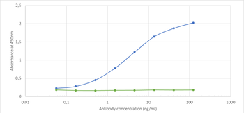
Binding curve of anti-CD155 antibody 3F1 (orb758858) to recombinant mouse CD155 Fc-Fusion Protein. ELISA Plate coated with recombinant mouse CD155 Fc-Fusion Protein at a concentration of 5 µg/ml. A 3-fold serial dilution from 10000 ng/ml was performed using orb758858. For detection, a 1:4000 dilution of HRP-labelled anti-rabbit antibody was used.
Immunofluorescence staining of RAW 264.7 cells using anti-CD155 antibody 3F1. Immunofluorescence analysis of paraformaldehyde fixed RAW 264.7 cells stained with the chimeric rabbit IgG version of 3F1 (orb758858) at 10 µg/ml followed by Alexa Fluor 488 secondary antibody (2 µg/ml), showing membrane staining. The nuclear stain is DAPI (blue). Panels show from left-right, top-bottom orb758858, DAPI, merged channels and an isotype control. The isotype control was stained with anti-Fluorescein antibody (orb256390) followed by Alexa Fluor 488 secondary antibody.
Western Blot using anti-CD155 antibody 3F1. Mouse thymus lysate (35 µg protein in RIPA buffer) was resolved on an SDS PAGE gel and blots probed with the chimeric rabbit IgG version of 3F1 (orb758858) at 0.1 µg/ml before detection using an anti-rabbit secondary antibody. A primary incubation of 1h was used and protein was detected by chemiluminescence.
* VAT and and shipping costs not included. Errors and price changes excepted