The original antibody was generated by cloning the variable regions of the scFvs selected from phage display libraries into separate vectors for IgG1 heavy-chain and light-chain expression. The harvested supernatents were then purified on protein A columns. The original immunogen was the whole irradiated virion.
Conjugation:
Unconjugated
Alternative Names:
NP, NC, Protein N, Nucleocapsid protein, SARS-CoV Protein N, SARS-CoV Nucleocapsid protein, SARS Coronavirus, SARS-CoV-2, SARS CoV 2, 2019-nCoV, sars-cov
Human monoclonal antibody to Covid-19 & SARS-CoV Nucleoprotein
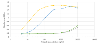
Binding curve of anti-SARS-Cov-2 (COVID-19) & SARS-CoV Nucleoprotein antibody CR3018 (03-018) (orb669769) to SARS-CoV-2 Nucleoproteins (REC31812 and REC31851). ELISA Plate coated with SARS-CoV-2 Nucleoproteins (REC31812/REC31851, Native Antigen Company) at a concentration of 5 µg/ml. A 3-fold serial dilution from 10000 ng/ml was performed using orb669769. For detection, a 1:4000 dilution of HRP-labelled anti-rabbit antibody was used. Rabbit anti-Fluorescein [4-4-20 (enhanced)] antibody (orb256390) was used as a control.
Immunofluorescence staining of MDCK-SIAT1 cells transfected with SARS-CoV-2 NP with anti-Covid-19 & SARS-CoV Nucleoprotein antibody CR3018 (03-018). Immunofluorescence analysis of MDCK-SIAT1 cells stably transfected with SARS-CoV-2 NP. The cells were seeded in a flat bottomed 96 well plate overnight, fixed in 10% formalin at 4C for 30 min, permeabilised for 20 min at RT and then stained with the human IgG1 version of CR3018 (03-018) (orb669768) in PBS/0.1% BSA at 10 µg/ml for 1 hour followed by a goat anti-human Alexa Fluor 647 (Invitrogen) secondary antibody.
* VAT and and shipping costs not included. Errors and price changes excepted