Each vial contains 4mg Trehalose, 0.9mg NaCl, 0.2mg Na2HPO4.
Form:
Lyophilized
Target:
Basigin
Application Dilute:
Western blot, 0.1-0.25µg/ml, Mouse, Rat Immunohistochemistry (Paraffin-embedded Section), 2-5µg/ml, Mouse, Rat Immunofluorescence, 5µg/ml, Mouse, Rat Flow Cytometry (Fixed), 1-3µg/1x10 6 cells, Mouse ELISA, 0.1-0.5µg/ml, -
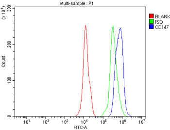
Flow Cytometry analysis of mouse PBMC cells using anti-CD147/Bsg antibody. Overlay histogram showing mouse PBMC cells (Blue line). The cells were fixed with 4% paraformaldehyde and blocked with 10% normal goat serum. And then incubated with rabbit anti-CD147/Bsg Antibody (1 µg/1x10 6 cells) for 30 min at 20C. DyLight488 conjugated goat anti-rabbit IgG (5-10 µg/1x10 6 cells) was used as secondary antibody for 30 minutes at 20C. Isotype control antibody (Green line) was rabbit IgG (1 µg/1x10 6) used under the same conditions. Unlabelled sample without incubation with primary antibody and secondary antibody (Red line) was used as a blank control.
IF analysis of CD147/Bsg using anti-CD147/Bsg antibody. CD147/Bsg was detected in a paraffin-embedded section of mouse colon tissue. Heat mediated antigen retrieval was performed in EDTA buffer (pH8.0, epitope retrieval solution). The tissue section was blocked with 10% goat serum. The tissue section was then incubated with 4 µg/mL rabbit anti-CD147/Bsg Antibody overnight at 4C. Biotin conjugated goat anti-rabbit IgG was used as secondary antibody and incubated for 30 minutes at 37C. The tissue section was developed using DyLight488 Conjugated Avidin. The section was counterstained with DAPI. Visualize using a fluorescence microscope and filter sets appropriate for the label used.
IF analysis of CD147/Bsg using anti-CD147/Bsg antibody. CD147/Bsg was detected in a paraffin-embedded section of rat colon tissue. Heat mediated antigen retrieval was performed in EDTA buffer (pH8.0, epitope retrieval solution). The tissue section was blocked with 10% goat serum. The tissue section was then incubated with 4 µg/mL rabbit anti-CD147/Bsg Antibody overnight at 4C. Biotin conjugated goat anti-rabbit IgG was used as secondary antibody and incubated for 30 minutes at 37C. The tissue section was developed using DyLight488 Conjugated Avidin. The section was counterstained with DAPI. Visualize using a fluorescence microscope and filter sets appropriate for the label used.
IHC analysis of CD147/Bsg using anti-CD147/Bsg antibody. CD147/Bsg was detected in a paraffin-embedded section of mouse colon tissue. Heat mediated antigen retrieval was performed in EDTA buffer (pH8.0, epitope retrieval solution). The tissue section was blocked with 10% goat serum. The tissue section was then incubated with 2 µg/ml rabbit anti-CD147/Bsg Antibody overnight at 4C. Biotinylated goat anti-rabbit IgG was used as secondary antibody and incubated for 30 minutes at 37C. The tissue section was developed using Strepavidin-Biotin-Complex (SABC) with DAB as the chromogen.
IHC analysis of CD147/Bsg using anti-CD147/Bsg antibody. CD147/Bsg was detected in a paraffin-embedded section of mouse kidney tissue. Heat mediated antigen retrieval was performed in EDTA buffer (pH8.0, epitope retrieval solution). The tissue section was blocked with 10% goat serum. The tissue section was then incubated with 2 µg/ml rabbit anti-CD147/Bsg Antibody overnight at 4C. Biotinylated goat anti-rabbit IgG was used as secondary antibody and incubated for 30 minutes at 37C. The tissue section was developed using Strepavidin-Biotin-Complex (SABC) with DAB as the chromogen.
IHC analysis of CD147/Bsg using anti-CD147/Bsg antibody. CD147/Bsg was detected in a paraffin-embedded section of rat kidney tissue. Heat mediated antigen retrieval was performed in EDTA buffer (pH8.0, epitope retrieval solution). The tissue section was blocked with 10% goat serum. The tissue section was then incubated with 2 µg/ml rabbit anti-CD147/Bsg Antibody overnight at 4C. Biotinylated goat anti-rabbit IgG was used as secondary antibody and incubated for 30 minutes at 37C. The tissue section was developed using Strepavidin-Biotin-Complex (SABC) with DAB as the chromogen.
IHC analysis of CD147/Bsg using anti-CD147/Bsg antibody. CD147/Bsg was detected in a paraffin-embedded section of rat testis tissue. Heat mediated antigen retrieval was performed in EDTA buffer (pH8.
* VAT and and shipping costs not included. Errors and price changes excepted