A synthetic peptide corresponding to a sequence in the middle region of human Cytokeratin 5, different from the related mouse sequence by one amino acid, and identical to the related rat sequence.
Conjugation:
Unconjugated
Alternative Names:
Interferon gamma, IFN-gamma, IFNG
Anti-Cytokeratin 5 Antibody (monoclonal, 5D3F7). Tested in Flow Cytometry, IF, IHC, ICC, WB applications. This antibody reacts with Human.
Clonality:
Monoclonal
Concentration:
Adding 0.2 ml of distilled water will yield a concentration of 500 µg/ml.
Application Notes: Western blot, 0.25-0.5 µg/ml, Human Immunohistochemistry(Paraffin-embedded Section), 2-5 µg/ml, Human Immunocytochemistry/Immunofluorescence, 5 µg/ml, Human Immunofluorescence, 5 µg/ml, Human Flow Cytometry (Fixed), 1-3 µg/1x106 cells, Human. Adding 0.2 ml of distilled water will yield a concentration of 500 µg/ml
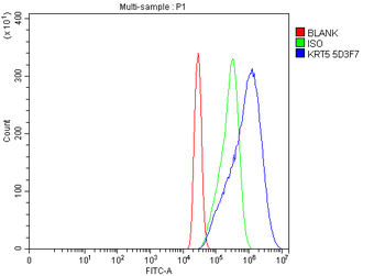
Flow Cytometry analysis of A431 cells using anti-Cytokeratin 5 antibody. Overlay histogram showing A431 cells (Blue line). To facilitate intracellular staining, cells were fixed with 4% paraformaldehyde and permeabilized with permeabilization buffer. The cells were blocked with 10% normal goat serum. And then incubated with mouse anti-Cytokeratin 5 Antibody (1 µg/1x10 6 cells) for 30 min at 20C. DyLight488 conjugated goat anti-mouse IgG (5-10 µg/1x10 6 cells) was used as secondary antibody for 30 minutes at 20C. Isotype control antibody (Green line) was mouse IgG (1 µg/1x10 6) used under the same conditions. Unlabelled sample without incubation with primary antibody and secondary antibody (Red line) was used as a blank control.
IF analysis of Cytokeratin 5 using anti-Cytokeratin 5 antibody. Cytokeratin 5 was detected in a paraffin-embedded section of human lung cancer tissue. Heat mediated antigen retrieval was performed in EDTA buffer (pH8.0, epitope retrieval solution). The tissue section was blocked with 10% goat serum. The tissue section was then incubated with 5 µg/mL mouse anti-Cytokeratin 5 Antibody overnight at 4C. Biotin conjugated goat anti-mouse IgG was used as secondary antibody and incubated for 30 minutes at 37C. The tissue section was developed using DyLight488 Conjugated Avidin. The section was counterstained with DAPI. Visualize using a fluorescence microscope and filter sets appropriate for the label used.
IF analysis of Cytokeratin 5 using anti-Cytokeratin 5 antibody. Cytokeratin 5 was detected in an immunocytochemical section of A431 cells. Enzyme antigen retrieval was performed using IHC enzyme antigen retrieval reagent for 15 mins. The cells were blocked with 10% goat serum. And then incubated with 5 µg/mL mouse anti-Cytokeratin 5 Antibody overnight at 4C. DyLight488 Conjugated Goat Anti-Mouse IgG was used as secondary antibody at 1:100 dilution and incubated for 30 minutes at 37C. The section was counterstained with DAPI. Visualize using a fluorescence microscope and filter sets appropriate for the label used.
IHC analysis of Cytokeratin 5 using anti-Cytokeratin 5 antibody. Cytokeratin 5 was detected in a paraffin-embedded section of human lung cancer tissue. Heat mediated antigen retrieval was performed in EDTA buffer (pH8.0, epitope retrieval solution). The tissue section was blocked with 10% goat serum. The tissue section was then incubated with 2 µg/ml mouse anti-Cytokeratin 5 Antibody overnight at 4C. Biotinylated goat anti-mouse IgG was used as secondary antibody and incubated for 30 minutes at 37C. The tissue section was developed using Strepavidin-Biotin-Complex (SABC) with DAB as the chromogen.
IHC analysis of Cytokeratin 5 using anti-Cytokeratin 5 antibody. Cytokeratin 5 was detected in a paraffin-embedded section of human metaplasia of squamous cells of the renal pelvis tissue. Heat mediated antigen retrieval was performed in EDTA buffer (pH8.0, epitope retrieval solution). The tissue section was blocked with 10% goat serum. The tissue section was then incubated with 2 µg/ml mouse anti-Cytokeratin 5 Antibody overnight at 4C. Biotinylated goat anti-mouse IgG was used as secondary antibody and incubated for 30 minutes at 37C. The tissue section was developed using Strepavidin-Biotin-Complex (SABC) with DAB as the chromogen.
IHC analysis of Cytokeratin 5 using anti-Cytokeratin 5 antibody. Cytokeratin 5 was detected in a paraffin-embedded section of human rectal moderately differentiated adenocarcinoma tissue. Heat mediated antigen retrieval was performed in EDTA buffer (pH8.0, epitope retrieval solution). The tissue section was blocked with 10% goat serum. The tissue section was then incubated with 2 µg/ml mouse anti-Cytokeratin 5 Antibody overnight at 4C. Biotinylated goat anti-mouse IgG was used as secondary antibody and incubated for 30 minutes at 37C. The tissue section was developed using Strepavidin-Biotin-Complex (SABC) with DAB as the chromogen.
Western blot analysis of Cy
* VAT and and shipping costs not included. Errors and price changes excepted