
ACTB Monoclonal Antibody, IgG2b, Clone: [1D12E12B5], Unconjugated, Mouse
Catalog Number:
CSB-MA000091M1M
- Images (7)
| Article Name: | ACTB Monoclonal Antibody, IgG2b, Clone: [1D12E12B5], Unconjugated, Mouse |
| Biozol Catalog Number: | CSB-MA000091M1M |
| Supplier Catalog Number: | CSB-MA000091M1m |
| Alternative Catalog Number: | CSB-MA000091M1M-100UL, CSB-MA000091M1M-50UL |
| Manufacturer: | Cusabio |
| Host: | Mouse |
| Category: | Antikörper |
| Application: | ELISA, FC, IF, IHC, WB |
| Species Reactivity: | Human, Mouse, Rabbit, Rat |
| Conjugation: | Unconjugated |
| Alternative Names: | ACTB |
| Clonality: | Monoclonal |
| Clone Designation: | [1D12E12B5] |
| Isotype: | IgG2b |
| Buffer: | Preservative: 0.03% Proclin 300<br />Constituents: 50% Glycerol, 0.01M PBS, PH 7.4 |
| Purity: | >95%, Protein A purified |
| Form: | Liquid |
| Target: | ACTB |
| Application Dilute: | Recommended dilution: WB: 1:5000-1:80000, IHC: 1:500-1:1000, IF: 1:50-1:200, FC: 1:100-1:300 |
|
|
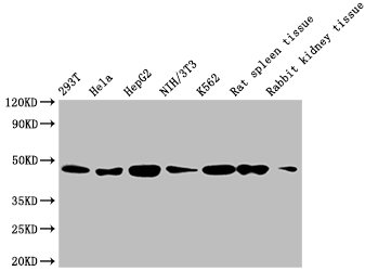
Western Blot Positive WB detected in: 293T whole cell lysate, Hela whole cell lysate, HepG2 whole cell lysate, NIH/3T3 whole cell lysate, K562 whole cell lysate, Rat spleen tissue, Rabbit kidney tissue All lanes: ACTB antibody at 1:5000 Secondary Goat polyclonal to mouse IgG at 1/50000 dilution Predicted band size: 42 KDa Observed band size: 42 KDa Exposure time: 5min |
|
|
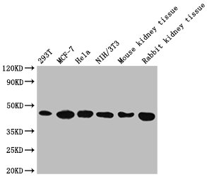
Western Blot Positive WB detected in: 293T whole cell lysate, MCF-7 whole cell lysate, Hela whole cell lysate, NIH/3T3 whole cell lysate, Mouse kidney tissue, Rabbit kidney tissue All lanes: ACTB antibody at 1:5000 Secondary Goat polyclonal to mouse IgG at 1/50000 dilution Predicted band size: 42 KDa Observed band size: 42 KDa Exposure time: 5min |
|
|
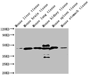
Western Blot Positive WB detected in: Mouse liver tissue, Mouse brain tissue, Mouse lung tissue, Mouse kidney tissue, Mouse spleen tissue, Mouse stomach tissue All lanes: ACTB antibody at 1:5000 Secondary Goat polyclonal to mouse IgG at 1/50000 dilution Predicted band size: 42 KDa Observed band size: 42 KDa Exposure time: 5min |
|
|
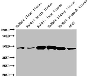
Western Blot Positive WB detected in: Rabbit liver tissue, Rabbit brain tissue, Rabbit lung tissue, Rabbit kidney tissue, Rabbit stomach tissue, A549 whole cell lysate All lanes: ACTB antibody at 1:5000 Secondary Goat polyclonal to mouse IgG at 1/50000 dilution Predicted band size: 42 KDa Observed band size: 42 KDa Exposure time: 5min |
|
|
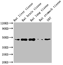
Western Blot Positive WB detected in: Rat liver tissue, Rat brain tissue, Rat lung tissue, Rat stomach tissue, U87 whole cell lysate All lanes: ACTB antibody at 1:5000 Secondary Goat polyclonal to mouse IgG at 1/50000 dilution Predicted band size: 42 KDa Observed band size: 42 KDa Exposure time: 5min |
|
|
CSB-MA000091M1m |
|
|
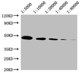
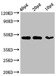
Western Blot Positive WB detected in: 293T whole cell lysate at 40ug, 20ug, 10ug All lanes:ACTB antibody at 1:5000 Secondary Goat polyclonal to mouse IgG at 1/50000 dilution Predicted band size: 42 KDa Observed band size: 42 KDa Exposure time: 5min |
Product Guarantee and Expert Support
