
FLOT1 Monoclonal Antibody, IgG2a, Clone: [4C8B6], Unconjugated, Mouse
Catalog Number:
CSB-MA008727A0M
- Images (5)

| Article Name: | FLOT1 Monoclonal Antibody, IgG2a, Clone: [4C8B6], Unconjugated, Mouse |
| Biozol Catalog Number: | CSB-MA008727A0M |
| Supplier Catalog Number: | CSB-MA008727A0m |
| Alternative Catalog Number: | CSB-MA008727A0M-100UL, CSB-MA008727A0M-50UL |
| Manufacturer: | Cusabio |
| Host: | Mouse |
| Category: | Antikörper |
| Application: | ELISA, FC, IF, IHC, WB |
| Species Reactivity: | Human, Mouse |
| Conjugation: | Unconjugated |
| Alternative Names: | FLOT 1 antibody, FLOT1 antibody, FLOT1_HUMAN antibody, Flotillin-1 antibody, Flotillin1 antibody, Integral membrane component of caveolae antibody, Reggie 2 antibody |
| Clonality: | Monoclonal |
| Clone Designation: | [4C8B6] |
| Isotype: | IgG2a |
| UniProt: | O75955 |
| Buffer: | PBS with 0.1% Sodium Azide, 50% Glycerol, pH 7.3. -20C, Avoid freeze / thaw cycles. |
| Purity: | >95%, Protein A purified |
| Form: | Liquid |
| Target: | FLOT1 |
| Application Dilute: | Recommended dilution: WB:1:1000-1:5000, IHC:1:50-1:200, IF:1:50-1:200, FC:1:50-1:200 |
|
|
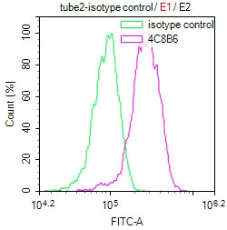
Overlay Peak curve showing Hela cells stained with CSB-MA008727A0m (red line) at 1:100. The cells were incubated in 10% normal goat serum to block non-specific protein-protein interactions followed by the antibody (1µg/1*106 cells) for 1h at 4C. The secondary antibody used was FITC-conjugated Goat Anti-Mouse IgG(H+L) at 1/100 dilution for 30min at 4C. Isotype control antibody (green line) was mouse IgG1 (1µg/1*106 cells) used under the same conditions. Acquisition of >10,000 events was performed. |
|
|
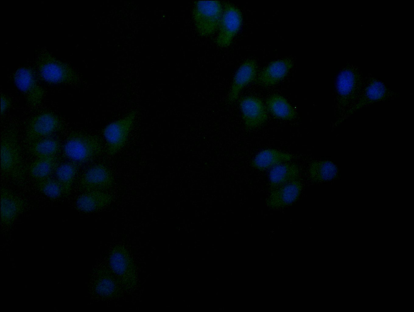
Immunofluorescence staining of Hela cells with CSB-MA008727A0m at 1:100, counter-stained with DAPI. The cells were fixed in 4% formaldehyde and blocked in 10% normal Goat Serum. The cells were incubated with the antibody overnight at 4C. Nuclear DNA was labeled in blue with DAPI. The secondary antibody was FITC-conjugated AffiniPure Goat Anti-Mouse IgG (H+L). |
|
|
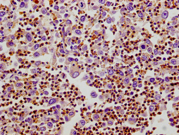
IHC image of CSB-MA008727A0m diluted at 1:100 and staining in paraffin-embedded human lung cancer tissue performed on a Leica BondTM system. After dewaxing and hydration, antigen retrieval was mediated by high pressure in a citrate buffer (pH 6.0). Section was blocked with 10% normal goat serum 30min at RT. Then primary antibody (1% BSA) was incubated at 4C overnight. The primary is detected by a biotinylated secondary antibody and visualized using an HRP conjugated SP system. |
|
|
IHC image of CSB-MA008727A0m diluted at 1:100 and staining in paraffin-embedded human cervical cancer tissue performed on a Leica BondTM system. After dewaxing and hydration, antigen retrieval was mediated by high pressure in a citrate buffer (pH 6.0). Section was blocked with 10% normal goat serum 30min at RT. Then primary antibody (1% BSA) was incubated at 4C overnight. The primary is detected by a biotinylated secondary antibody and visualized using an HRP conjugated SP system. |
|
|
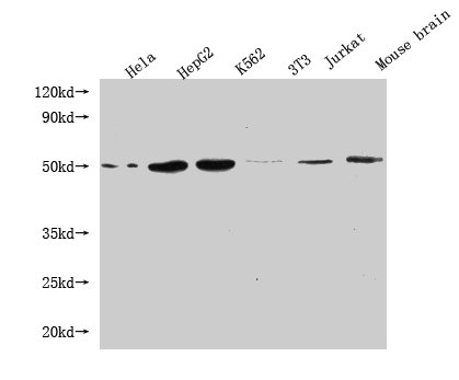
Western Blot Positive WB detected in: Hela whole cell lysate, HepG2 whole cell lysate, K562 whole cell lysate, 3T3 whole cell lysate, Jurkat whole cell lysate, Mouse brain tissue All lanes: FLOT1 antibody at 1:1000 Secondary Goat polyclonal to mouse IgG at 1/50000 dilution Predicted band size: 48, 43 kDa Observed band size: 48 KDa Exposure time:5min |
Product Guarantee and Expert Support
