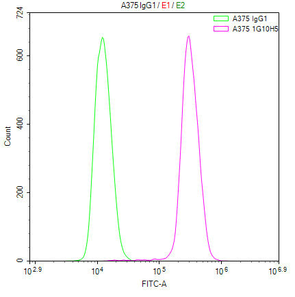
Overlay Peak curve showing A375 cells stained with CSB-MA013331A0m (red line) at 1:100. Then 10% normal goat serum was Incubated to block non-specific protein-protein interactions followed by the antibody (1µg/1*106cells) for 45 min at 4C. The secondary antibody used was FITC-conjugated Goat Anti-Mouse IgG(H+L) at 1/200 dilution for 35 min at 4C. Isotype control antibody (green line) was mouse IgG1 (1µg/1*106cells) used under the same conditions. Acquisition of >10,000 events was performed.
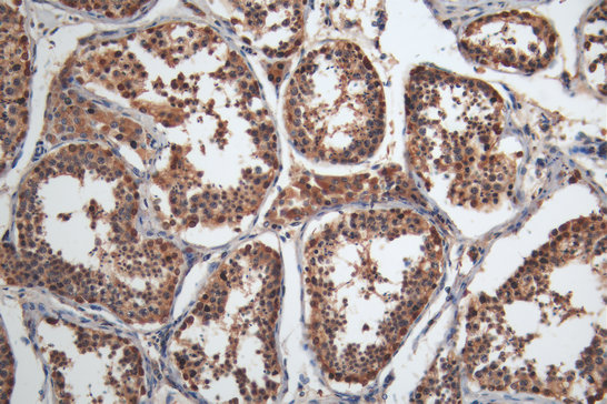
IHC image of CSB-MA013330A0m diluted at 1:200 and staining in paraffin-embedded human testis tissue performed on a Leica BondTM system. After dewaxing and hydration, antigen retrieval was mediated by high pressure in a citrate buffer (pH 6.0). Section was blocked with 10% normal goat serum 30min at RT. Then primary antibody (1% BSA) was incubated at 4C overnight. The primary is detected by a Goat anti-mouse polymer IgG labeled by HRP and visualized using 0.05% DAB.
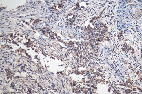
IHC image of CSB-MA013330A0m diluted at 1:200 and staining in paraffin-embedded human bladder cancer performed on a Leica BondTM system. After dewaxing and hydration, antigen retrieval was mediated by high pressure in a citrate buffer (pH 6.0). Section was blocked with 10% normal goat serum 30min at RT. Then primary antibody (1% BSA) was incubated at 4C overnight. The primary is detected by a Goat anti-mouse polymer IgG labeled by HRP and visualized using 0.05% DAB.
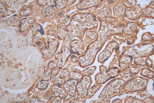
IHC image of CSB-MA013330A0m diluted at 1:200 and staining in paraffin-embedded human placenta tissue performed on a Leica BondTM system. After dewaxing and hydration, antigen retrieval was mediated by high pressure in a citrate buffer (pH 6.0). Section was blocked with 10% normal goat serum 30min at RT. Then primary antibody (1% BSA) was incubated at 4C overnight. The primary is detected by a Goat anti-mouse polymer IgG labeled by HRP and visualized using 0.05% DAB.
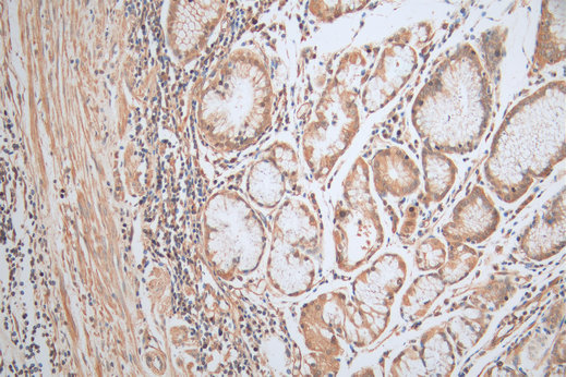
IHC image of CSB-MA013330A0m diluted at 1:200 and staining in paraffin-embedded human gastric cancer tissue performed on a Leica BondTM system. After dewaxing and hydration, antigen retrieval was mediated by high pressure in a citrate buffer (pH 6.0). Section was blocked with 10% normal goat serum 30min at RT. Then primary antibody (1% BSA) was incubated at 4C overnight. The primary is detected by a Goat anti-mouse polymer IgG labeled by HRP and visualized using 0.05% DAB.
* VAT and and shipping costs not included. Errors and price changes excepted