
Fxn Antibody, Unconjugated, Rabbit, Polyclonal
Catalog Number:
CSB-PA009086LA01MO
- Images (6)

| Article Name: | Fxn Antibody, Unconjugated, Rabbit, Polyclonal |
| Biozol Catalog Number: | CSB-PA009086LA01MO |
| Supplier Catalog Number: | CSB-PA009086LA01MO |
| Alternative Catalog Number: | CSB-PA009086LA01MO-100UL |
| Manufacturer: | Cusabio |
| Host: | Rabbit |
| Category: | Antikörper |
| Application: | ELISA, IHC, WB |
| Species Reactivity: | Human, Mouse |
| Conjugation: | Unconjugated |
| Alternative Names: | Fxn antibody, FrdaFrataxin antibody, mitochondrial antibody, Fxn antibody, EC 1.16.3.1) [Cleaved into: Frataxin intermediate form, Frataxin mature form] antibody |
| Clonality: | Polyclonal |
| UniProt: | O35943 |
| Buffer: | Preservative: 0.02% sodium azide<br />Constituents: 50% Glycerol, 0.01M PBS, pH 7.4 |
| Purity: | Antigen Affinity Purified |
| Form: | Liquid |
| Target: | Fxn |
| Application Dilute: | Recommended dilution: WB:1:500-1:2000, IHC:1:50-1:200 |
|
|
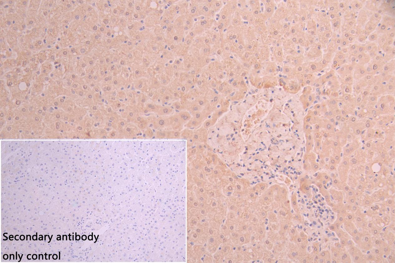
IHC image of CSB-PA009086LA01MO diluted at 1:66 and staining in paraffin-embedded human Liver tissue performed on a Leica BondTM system. After dewaxing and hydration, antigen retrieval was mediated by high pressure in a citrate buffer (pH 6.0). Section was blocked with 10% normal goat serum 30min at RT. Then primary antibody (1% BSA) was incubated at 4C overnight. The primary is detected by a Goat anti-rabbit polymer IgG labeled by HRP and visualized using 0.05% DAB. Secondary antibody only control: uses 1% BSA instead of primary antibody |
|
|
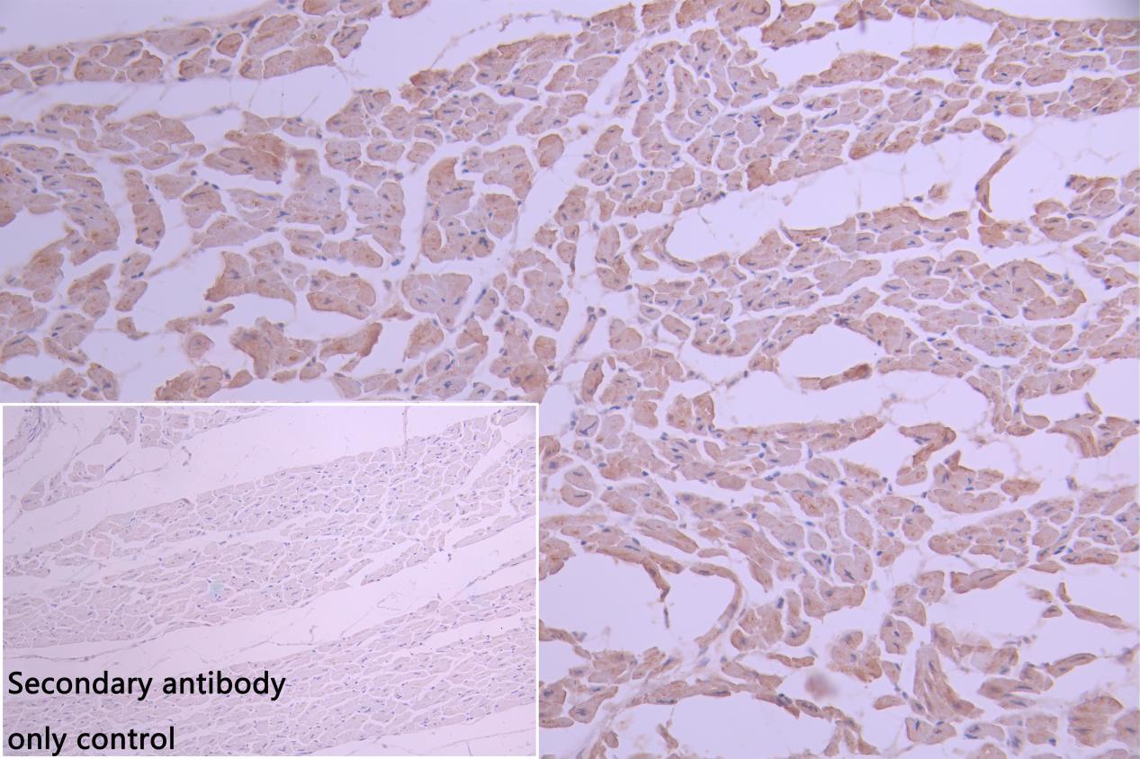
IHC image of CSB-PA009086LA01MO diluted at 1:66 and staining in paraffin-embedded human Heart tissue performed on a Leica BondTM system. After dewaxing and hydration, antigen retrieval was mediated by high pressure in a citrate buffer (pH 6.0). Section was blocked with 10% normal goat serum 30min at RT. Then primary antibody (1% BSA) was incubated at 4C overnight. The primary is detected by a Goat anti-rabbit polymer IgG labeled by HRP and visualized using 0.05% DAB. Secondary antibody only control: uses 1% BSA instead of primary antibody |
|
|
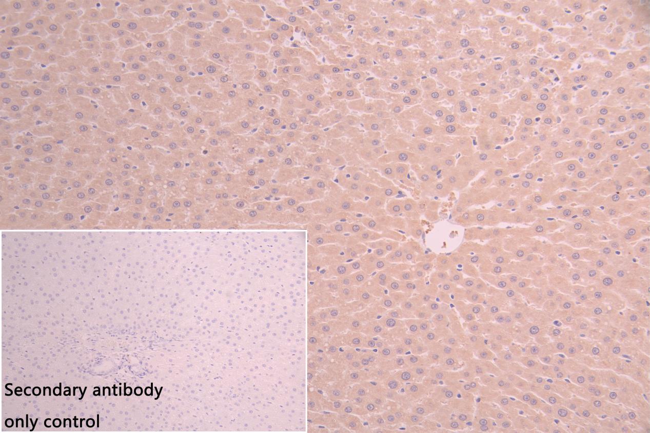
IHC image of CSB-PA009086LA01MO diluted at 1:66 and staining in paraffin-embedded rat Liver tissue performed on a Leica BondTM system. After dewaxing and hydration, antigen retrieval was mediated by high pressure in a citrate buffer (pH 6.0). Section was blocked with 10% normal goat serum 30min at RT. Then primary antibody (1% BSA) was incubated at 4C overnight. The primary is detected by a Goat anti-rabbit polymer IgG labeled by HRP and visualized using 0.05% DAB. Secondary antibody only control: uses 1% BSA instead of primary antibody |
|
|
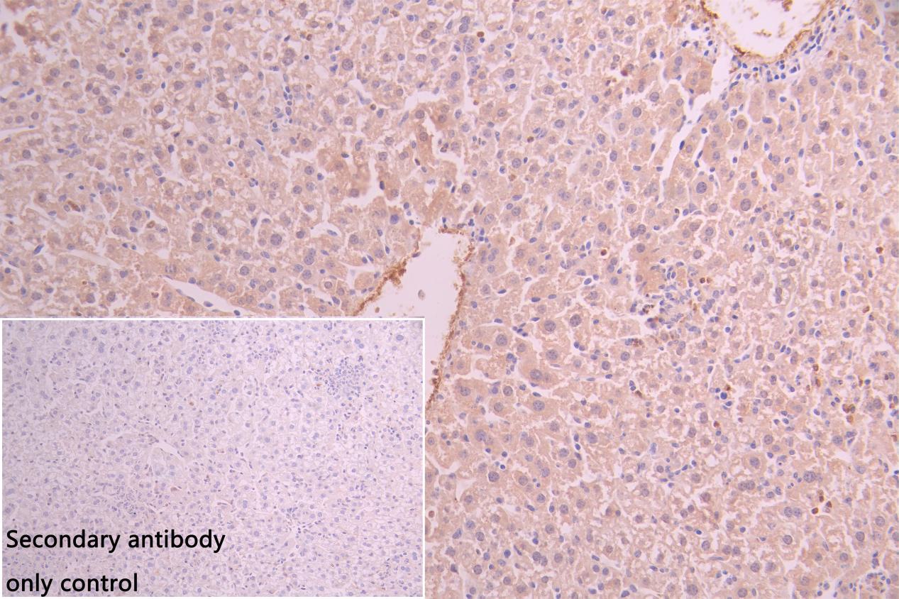
IHC image of CSB-PA009086LA01MO diluted at 1:66 and staining in paraffin-embedded mouse Liver tissue performed on a Leica BondTM system. After dewaxing and hydration, antigen retrieval was mediated by high pressure in a citrate buffer (pH 6.0). Section was blocked with 10% normal goat serum 30min at RT. Then primary antibody (1% BSA) was incubated at 4C overnight. The primary is detected by a Goat anti-rabbit polymer IgG labeled by HRP and visualized using 0.05% DAB. Secondary antibody only control: uses 1% BSA instead of primary antibody |
|
|
IHC image of CSB-PA009086LA01MO diluted at 1:66 and staining in paraffin-embedded mouse Heart tissue performed on a Leica BondTM system. After dewaxing and hydration, antigen retrieval was mediated by high pressure in a citrate buffer (pH 6.0). Section was blocked with 10% normal goat serum 30min at RT. Then primary antibody (1% BSA) was incubated at 4C overnight. The primary is detected by a Goat anti-rabbit polymer IgG labeled by HRP and visualized using 0.05% DAB. Secondary antibody only control: uses 1% BSA instead of primary antibody |
|
|
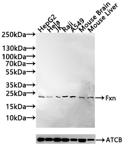
Western Blot Positive WB detected in: HepG2 whole cell lysate(30µg), Hela whole cell lysate(30µg), JK whole cell lysate(20µg), Raji whole cell lysate(30µg), A549 whole cell lysate(30µg), Mouse Brain tissue lysate(30µg),Mouse Liver tissue lysate(30µg) All lanes: Fxn antibody at 1:1000 Secondary Goat polyclonal to rabbit IgG at 1/20000 dilution Predicted band size: 23 kDa Observed band size: 23 kDa Exposure time: 120s |
Product Guarantee and Expert Support
