
KLRK1 Antibody, Unconjugated, Rabbit, Polyclonal
Catalog Number:
CSB-PA012474LA01HU
- Images (7)

| Article Name: | KLRK1 Antibody, Unconjugated, Rabbit, Polyclonal |
| Biozol Catalog Number: | CSB-PA012474LA01HU |
| Supplier Catalog Number: | CSB-PA012474LA01HU |
| Alternative Catalog Number: | CSB-PA012474LA01HU-100UL, CSB-PA012474LA01HU-50UL |
| Manufacturer: | Cusabio |
| Host: | Rabbit |
| Category: | Antikörper |
| Application: | ELISA, IF, IHC, WB |
| Species Reactivity: | Human |
| Conjugation: | Unconjugated |
| Alternative Names: | CD314 antibody, CD314 antigen antibody, D12S2489E antibody, Killer cell lectin like receptor subfamily K member 1 antibody, Killer cell lectin-like receptor subfamily K member 1 antibody, KLR antibody, KLRC4 KLRK1 readthrough antibody, KLRK1 antibody, NK cell receptor D antibody, NK lectin-like receptor antibody, NKG2 D activating NK receptor antibody, NKG2 D type II integral membrane protein antibody, NKG2-D antibody, NKG2-D type II integral membrane protein antibody, NKG2-D-activating NK receptor antibody, Nkg2d antibody, NKG2D_HUMAN antibody, NKLLR antibody, NKR P2 antibody, Nkrp2 antibody |
| Clonality: | Polyclonal |
| UniProt: | P26718 |
| Buffer: | pH7.4 PBS, 0.05% NaN3, 40% Glycerol |
| Purity: | >95%, Protein G purified |
| Form: | Liquid |
| Target: | KLRK1 |
| Application Dilute: | Recommended dilution: WB:1:500-1:2000, IHC:1:50-1:200, IF:1:20-1:100 |
|
|
|
|
|
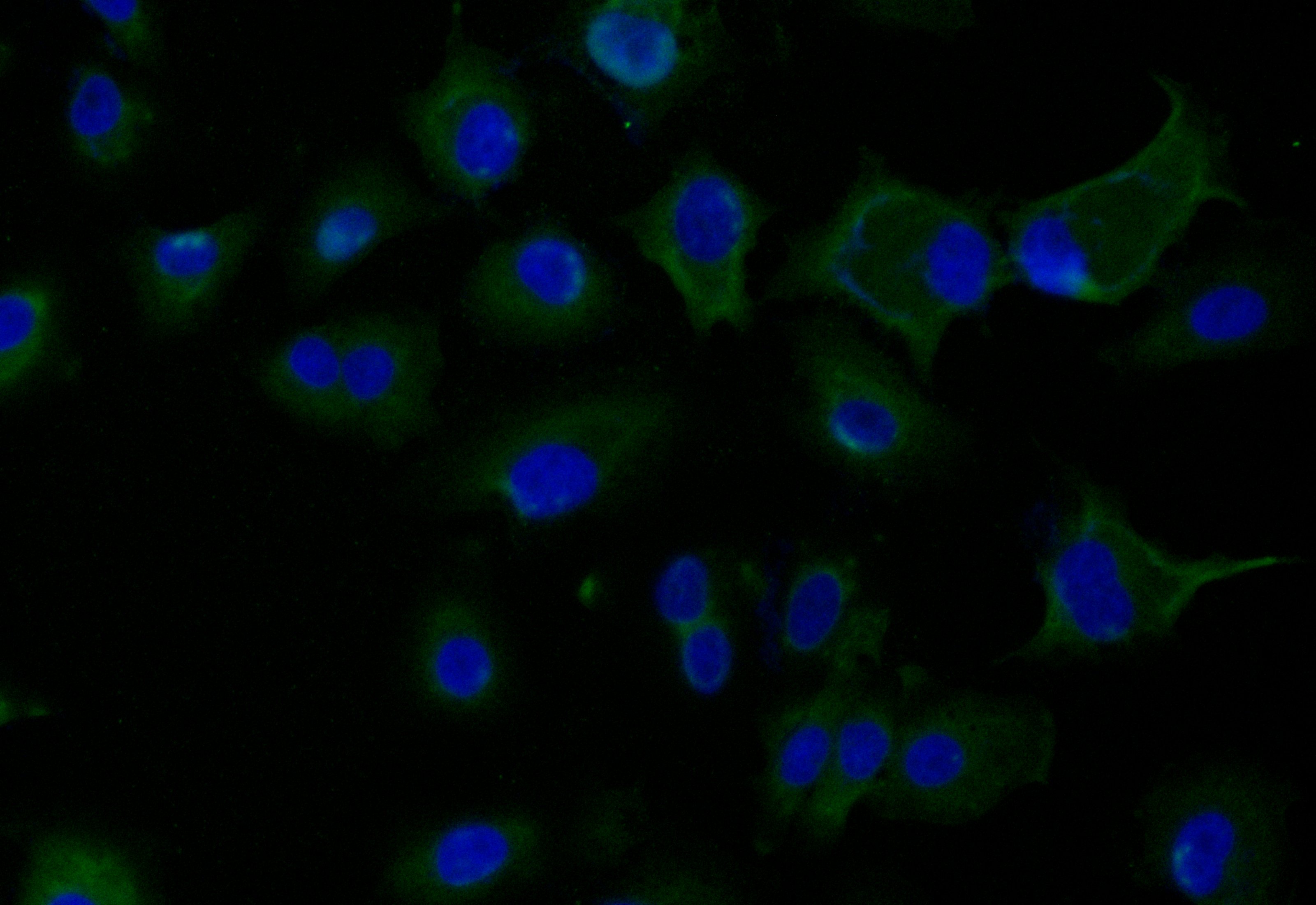
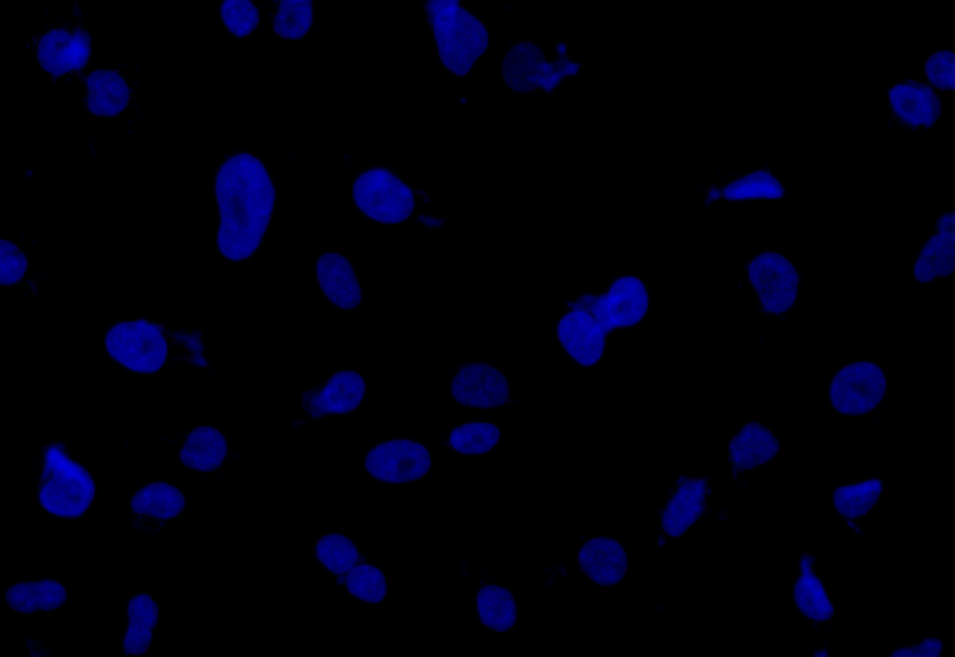
Immunofluorescence staining of Hela cell with CSB-PA012474LA01HU at 1:20, counter-stained with DAPI. The cells were fixed in 4% formaldehyde and blocked in 10% normal Goat Serum. The cells were then incubated with the antibody overnight at 4C. The secondary antibody was Alexa Fluor 488-congugated AffiniPure Goat Anti-Rabbit IgG(H+L). |
|
|
Immunofluorescence staining of Hela cell with 5% goat serum, counter-stained with DAPI. The cells were fixed in 4% formaldehyde and blocked in 10% normal Goat Serum. The cells were then incubated with the antibody overnight at 4C. The secondary antibody was Alexa Fluor 488-congugated AffiniPure Goat Anti-Rabbit IgG(H+L). |
|
|
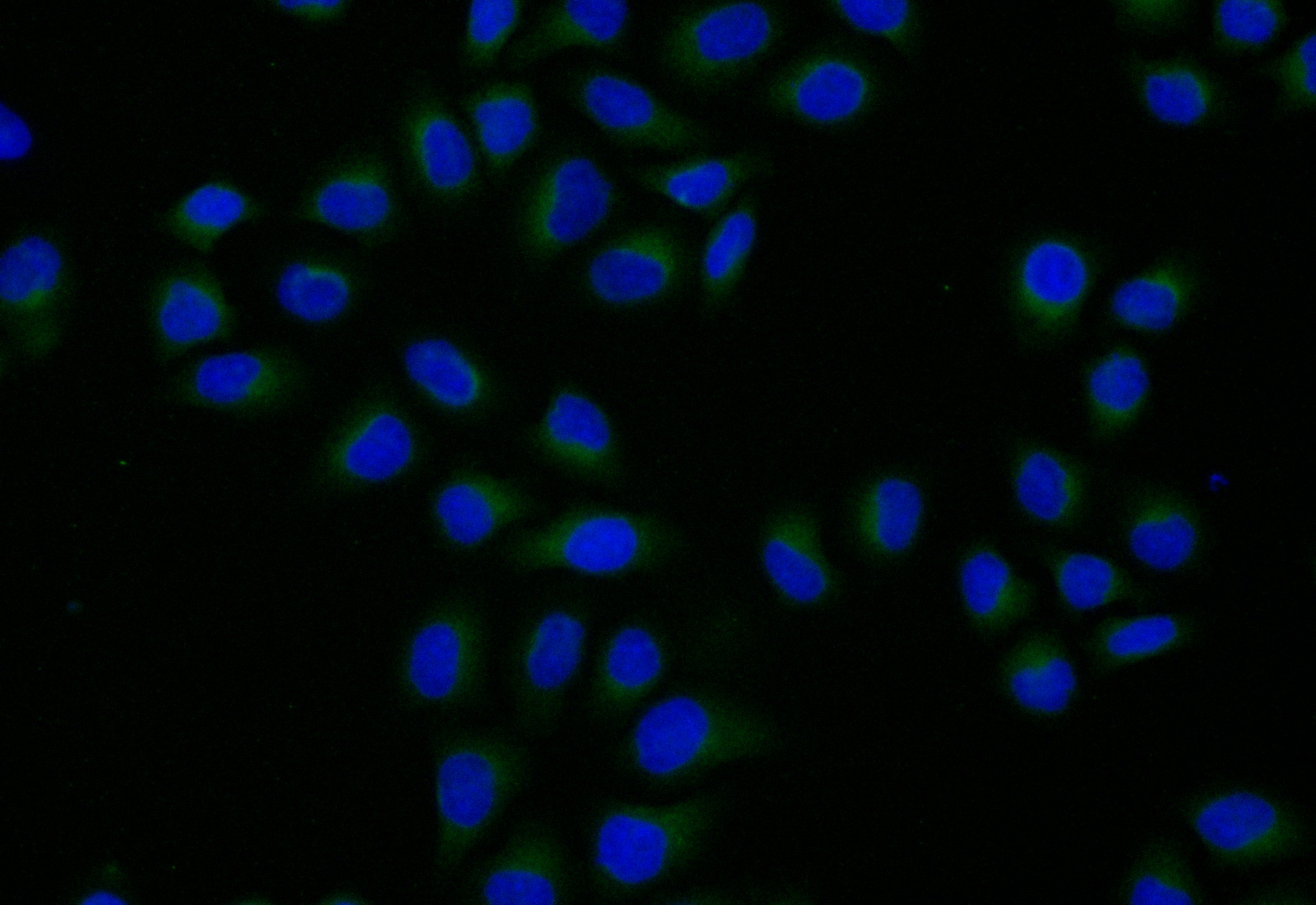
Immunofluorescence staining of A549 cell with CSB-PA012474LA01HU at 1:20, counter-stained with DAPI. The cells were fixed in 4% formaldehyde and blocked in 10% normal Goat Serum. The cells were then incubated with the antibody overnight at 4C. The secondary antibody was Alexa Fluor 488-congugated AffiniPure Goat Anti-Rabbit IgG(H+L). |
|
|
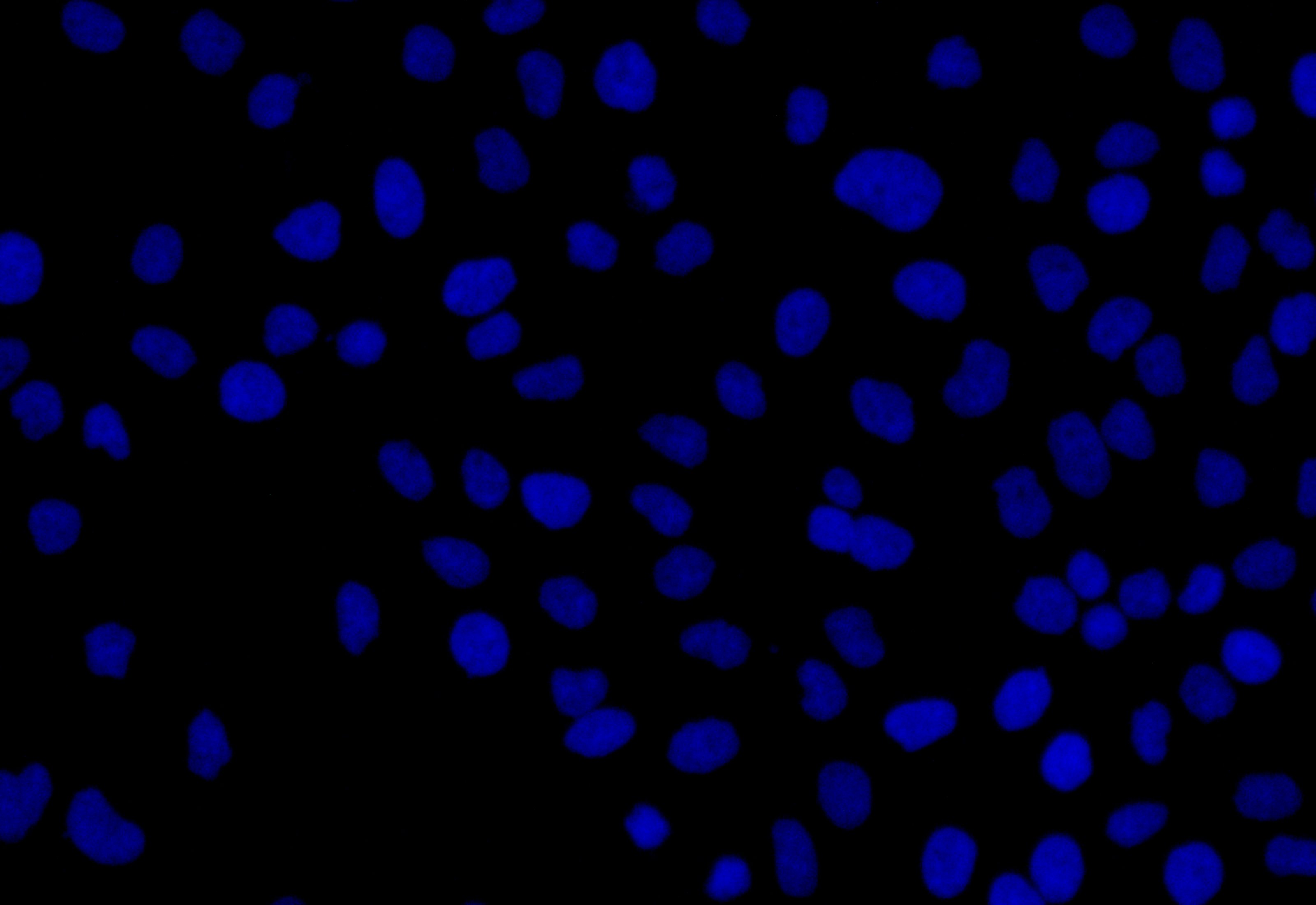
Immunofluorescence staining of A549 cell with 5% goat serum, counter-stained with DAPI. The cells were fixed in 4% formaldehyde and blocked in 10% normal Goat Serum. The cells were then incubated with the antibody overnight at 4C. The secondary antibody was Alexa Fluor 488-congugated AffiniPure Goat Anti-Rabbit IgG(H+L). |
|
|
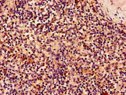
IHC image of CSB-PA012474LA01HU diluted at 1:50 and staining in paraffin-embedded human tonsil tissue performed on a Leica BondTM system. After dewaxing and hydration, antigen retrieval was mediated by high pressure in a citrate buffer (pH 6.0). Section was blocked with 10% normal goat serum 30min at RT. Then primary antibody (1% BSA) was incubated at 4C overnight. The primary is detected by a Goat anti-rabbit polymer IgG labeled by HRP and visualized using 0.05% DAB. Secondary antibody only control: uses 1% BSA instead of primary antibody |
|
|
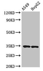
Western Blot Positive WB detected in: THP-1 whole cell lysate(30µg), A549 whole cell lysate(30µg), U251 whole cell lysate(30µg), Hela whole cell lysate(30µg) All lanes: KLRK1 antibody at 1:1000 Secondary Goat polyclonal to rabbit IgG at 1/20000 dilution Predicted band size: 26kDa Observed band size: 36 kDa Exposure time: 120s |
Product Guarantee and Expert Support
