
YWHAE Antibody, Unconjugated, Rabbit, Polyclonal
Catalog Number:
CSB-PA026287DA01HU
- Images (6)
| Article Name: | YWHAE Antibody, Unconjugated, Rabbit, Polyclonal |
| Biozol Catalog Number: | CSB-PA026287DA01HU |
| Supplier Catalog Number: | CSB-PA026287DA01HU |
| Alternative Catalog Number: | CSB-PA026287DA01HU-100UG, CSB-PA026287DA01HU-50UG |
| Manufacturer: | Cusabio |
| Host: | Rabbit |
| Category: | Antikörper |
| Application: | ELISA, IF, IHC, WB |
| Species Reactivity: | Human, Mouse |
| Conjugation: | Unconjugated |
| Alternative Names: | 14 3 3 E antibody, 14 3 3 epsilon antibody, 14 3 3E antibody, 14-3-3 protein epsilon antibody, 14-3-3E antibody, 1433E_HUMAN antibody, Epididymis luminal protein 2 antibody, FLJ45465 antibody, FLJ53559 antibody, HEL2 antibody, KCIP 1 antibody, KCIP1 antibody, MDCR antibody, MDS antibody, Mitochondrial import stimulation factor L subunit antibody, Protein kinase C inhibitor protein1 antibody, Tyrosine 3 monooxygenase/tryptophan 5 monooxygenase activation protein, epsilon antibody, Tyrosine 3 monooxygenase/tryptophan 5 monooxygenase activation protein, epsilon polypeptide antibody, Tyrosine 3/tryptophan 5 monooxygenase activation protein epsilon polypeptide antibody, YWHAE antibody |
| Clonality: | Polyclonal |
| UniProt: | P62258 |
| Buffer: | Preservative: 0.03% Proclin 300<br />Constituents: 50% Glycerol, 0.01M PBS, PH 7.4 |
| Purity: | >95%, Protein G purified |
| Form: | Liquid |
| Target: | YWHAE |
| Application Dilute: | Recommended dilution: WB:1:500-1:5000, IHC:1:1000-1:2000, IF:1:200-1:500 |
|
|
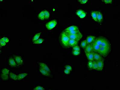
Immunofluorescence staining of HepG2 cells with CSB-PA026287DA01HU at 1:333, counter-stained with DAPI. The cells were fixed in 4% formaldehyde, permeabilized using 0.2% Triton X-100 and blocked in 10% normal Goat Serum. The cells were then incubated with the antibody overnight at 4°,C. The secondary antibody was Alexa Fluor 488-congugated AffiniPure Goat Anti-Rabbit IgG(H+L). |
|
|
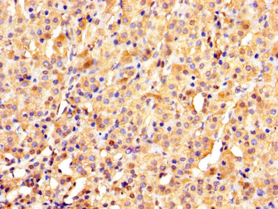
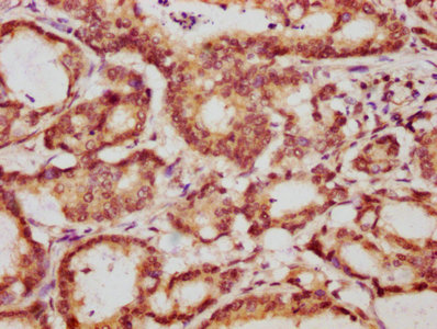
IHC image of CSB-PA026287DA01HU diluted at 1:1000 and staining in paraffin-embedded human adrenal gland tissue performed on a Leica BondTM system. After dewaxing and hydration, antigen retrieval was mediated by high pressure in a citrate buffer (pH 6.0). Section was blocked with 10% normal goat serum 30min at RT. Then primary antibody (1% BSA) was incubated at 4°,C overnight. The primary is detected by a biotinylated secondary antibody and visualized using an HRP conjugated SP system. |
|
|
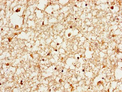
IHC image of CSB-PA026287DA01HU diluted at 1:1000 and staining in paraffin-embedded human brain tissue performed on a Leica BondTM system. After dewaxing and hydration, antigen retrieval was mediated by high pressure in a citrate buffer (pH 6.0). Section was blocked with 10% normal goat serum 30min at RT. Then primary antibody (1% BSA) was incubated at 4°,C overnight. The primary is detected by a biotinylated secondary antibody and visualized using an HRP conjugated SP system. |
|
|
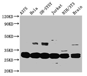
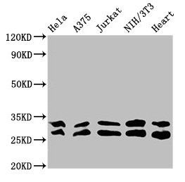
Western Blot Positive WB detected in: A375 whole cell lysate, Hela whole cell lysate, SH-SY5Y whole cell lysate, Jurkat whole cell lysate, NIH/3T3 whole cell lysate, Mouse brain tissue All lanes: YWHAE antibody at 3.3µg/ml Secondary Goat polyclonal to rabbit IgG at 1/50000 dilution Predicted band size: 30, 27 kDa Observed band size: 30 kDa |
|
|
IHC image of CSB-PA026287DA01HU diluted at 1:1000 and staining in paraffin-embedded human colon cancer performed on a Leica BondTM system. After dewaxing and hydration, antigen retrieval was mediated by high pressure in a citrate buffer (pH 6.0). Section was blocked with 10% normal goat serum 30min at RT. Then primary antibody (1% BSA) was incubated at 4°,C overnight. The primary is detected by a biotinylated secondary antibody and visualized using an HRP conjugated SP system. |
|
|
Western Blot Positive WB detected in: Hela whole cell lysate, A375 whole cell lysate, Jurkat whole cell lysate, NIH/3T3 whole cell lysate, Mouse heart tissue All lanes: YWHAE antibody at 3.3µg/ml Secondary Goat polyclonal to rabbit IgG at 1/50000 dilution Predicted band size: 30, 27 kDa Observed band size: 30, 27 kDa |
Product Guarantee and Expert Support
