
IFNA2 Antibody, Unconjugated, Rabbit, Polyclonal
Catalog Number:
CSB-PA06504A0RB
- Images (7)


| Article Name: | IFNA2 Antibody, Unconjugated, Rabbit, Polyclonal |
| Biozol Catalog Number: | CSB-PA06504A0RB |
| Supplier Catalog Number: | CSB-PA06504A0Rb |
| Alternative Catalog Number: | CSB-PA06504A0RB-100UL, CSB-PA06504A0RB-50UL |
| Manufacturer: | Cusabio |
| Host: | Rabbit |
| Category: | Antikörper |
| Application: | ELISA, IHC, WB |
| Species Reactivity: | Human, Mouse, Rat |
| Conjugation: | Unconjugated |
| Alternative Names: | Alpha 2a interferon antibody, IFN alpha 2b antibody, IFN-alpha-2 antibody, IFNA antibody, Ifna2 antibody, IFNA2_HUMAN antibody, INFA2 antibody, Interferon alpha 2 antibody, Interferon alpha A antibody, Interferon alpha-2 antibody, Interferon alpha-A antibody, LeIF A antibody, LeIFA antibody |
| Clonality: | Polyclonal |
| UniProt: | P01563 |
| Buffer: | Preservative: 0.05% NaN3<br /> Constituents: PBS containing 40% glycerol,pH7.4 |
| Purity: | Antigen Affinity Purified |
| Form: | Liquid |
| Target: | IFNA2 |
| Application Dilute: | Recommended dilution: WB:1:1000-1:5000 , IHC:1:100-1:300 |
|
|
Immunohistochemistry |
|
|
|
|
|
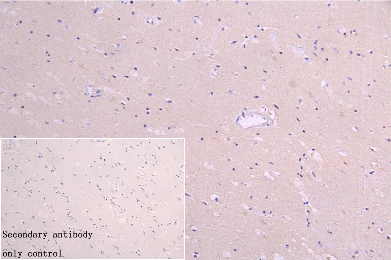
IHC image of CSB-PA06504A0Rb diluted at 1:200 and staining in paraffin-embedded human brain tissue performed on a Leica BondTM system. After dewaxing and hydration, antigen retrieval was mediated by high pressure in a citrate buffer (pH 6.0). Section was blocked with 10% normal goat serum 30min at RT. Then primary antibody (1% BSA) was incubated at 4C overnight. The primary is detected by a Goat anti-rabbit polymer IgG labeled by HRP and visualized using 0.05% DAB. |
|
|
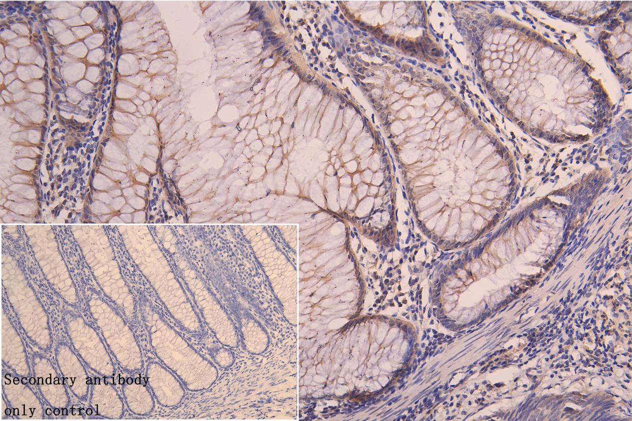
IHC image of CSB-PA06504A0Rb diluted at 1:200 and staining in paraffin-embedded human colorectal cancer performed on a Leica BondTM system. After dewaxing and hydration, antigen retrieval was mediated by high pressure in a citrate buffer (pH 6.0). Section was blocked with 10% normal goat serum 30min at RT. Then primary antibody (1% BSA) was incubated at 4C overnight. The primary is detected by a Goat anti-rabbit polymer IgG labeled by HRP and visualized using 0.05% DAB. |
|
|
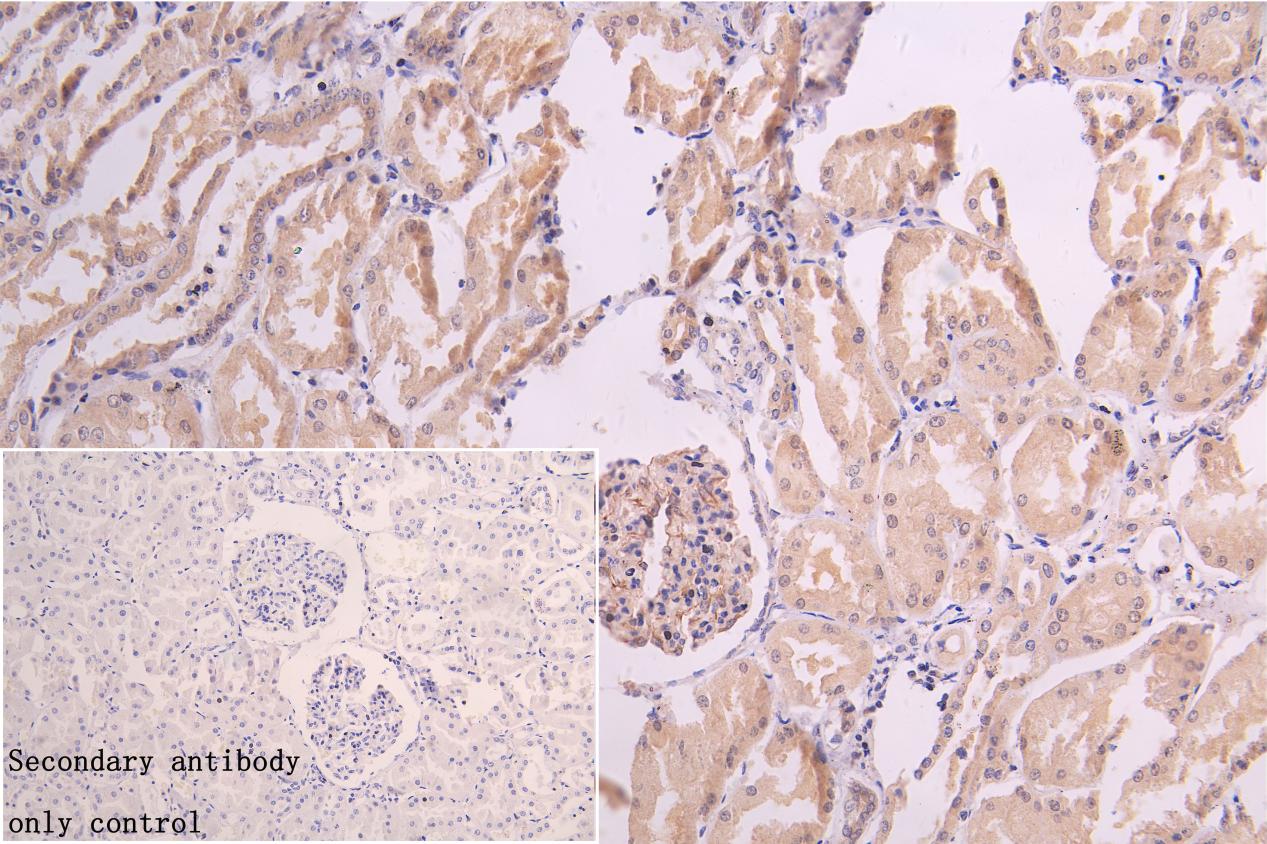
IHC image of CSB-PA06504A0Rb diluted at 1:200 and staining in paraffin-embedded human kidney tissue performed on a Leica BondTM system. After dewaxing and hydration, antigen retrieval was mediated by high pressure in a citrate buffer (pH 6.0). Section was blocked with 10% normal goat serum 30min at RT. Then primary antibody (1% BSA) was incubated at 4C overnight. The primary is detected by a Goat anti-rabbit polymer IgG labeled by HRP and visualized using 0.05% DAB. |
|
|
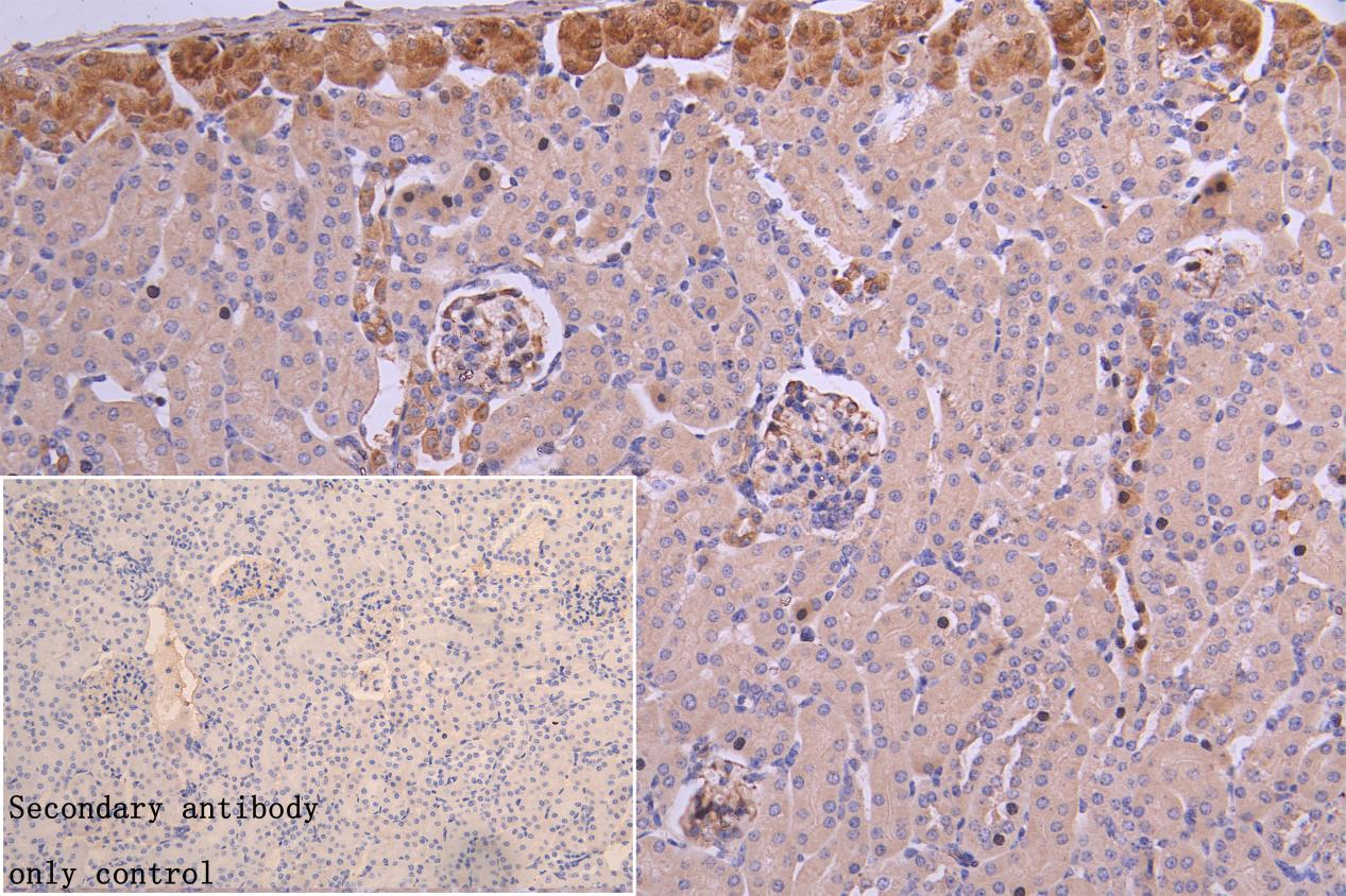
IHC image of CSB-PA06504A0Rb diluted at 1:200 and staining in paraffin-embedded Mouse kidney tissue performed on a Leica BondTM system. After dewaxing and hydration, antigen retrieval was mediated by high pressure in a citrate buffer (pH 6.0). Section was blocked with 10% normal goat serum 30min at RT. Then primary antibody (1% BSA) was incubated at 4C overnight. The primary is detected by a Goat anti-rabbit polymer IgG labeled by HRP and visualized using 0.05% DAB. |
|
|
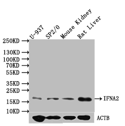
Western Blot Positive WB detected in: U-937 whole cell lysate, SP2/0 whole cell lysate, Mouse Kidney tissue lysate, Rat Liver tissue lysate All lanes: IFNA2 antibody at 1:1000 Secondary Goat polyclonal to rabbit IgG at 1/50000 dilution Predicted band size: 22 kDa Observed band size: 18-22 kDa |
Product Guarantee and Expert Support
