
CD9 Antibody, Unconjugated, Rabbit, Polyclonal
Catalog Number:
CSB-PA10559A0RB
- Images (7)


| Article Name: | CD9 Antibody, Unconjugated, Rabbit, Polyclonal |
| Biozol Catalog Number: | CSB-PA10559A0RB |
| Supplier Catalog Number: | CSB-PA10559A0Rb |
| Alternative Catalog Number: | CSB-PA10559A0RB-100UL |
| Manufacturer: | Cusabio |
| Host: | Rabbit |
| Category: | Antikörper |
| Application: | ELISA, IHC, WB |
| Species Reactivity: | Human |
| Conjugation: | Unconjugated |
| Alternative Names: | Tetraspanin 29 antibody, 5H9 antibody, 5H9 antigen antibody, Antigen defined by monoclonal antibody 602 29 antibody, Antigen defined by monoclonal antibody 60229 antibody, BA-2/p24 antigen antibody, BA2 antibody, BTCC 1 antibody, BTCC1 antibody, CD9 antibody, CD9 antigen antibody, CD9 antigen p24 antibody, CD9 molecule antibody, CD9_HUMAN antibody, Cell growth inhibiting gene 2 protein antibody, Cell growth-inhibiting gene 2 protein antibody, DRAP 27 antibody, DRAP27 antibody, GIG2 antibody, Growth inhibiting gene 2 protein antibody, Leukocyte antigen MIC3 antibody, MIC3 antibody, Motility related protein antibody, Motility-related protein antibody, MRP 1 antibody, MRP-1 antibody, MRP1 antibody, p24 antibody, p24 antigen antibody, Tetraspanin-29 antibody, Tspan 29 antibody, Tspan-29 antibody, TSPAN29 antibody |
| Clonality: | Polyclonal |
| UniProt: | P21926 |
| Buffer: | PBS with 0.1% Sodium Azide, 50% Glycerol, pH 7.3. -20C, Avoid freeze / thaw cycles. |
| Purity: | Antigen Affinity Purified |
| Form: | Liquid |
| Target: | CD9 |
| Application Dilute: | Recommended dilution: WB:1:500-1:3000, IHC:1:20-1:200 |
|
|
|
|
|
IHC image of CSB-PA10559A0Rb diluted at 1:50 and staining in paraffin-embedded human small tonsil tissue performed on a Leica BondTM system. After dewaxing and hydration, antigen retrieval was mediated by high pressure in a citrate buffer (pH 6.0). Section was blocked with 10% normal goat serum 30min at RT. Then primary antibody (1% BSA) was incubated at 4C overnight. The primary is detected by a Goat anti-rabbit polymer IgG labeled by HRP and visualized using 0.05% DAB. Secondary antibody only control: uses 1% BSA instead of primary antibody |
|
|
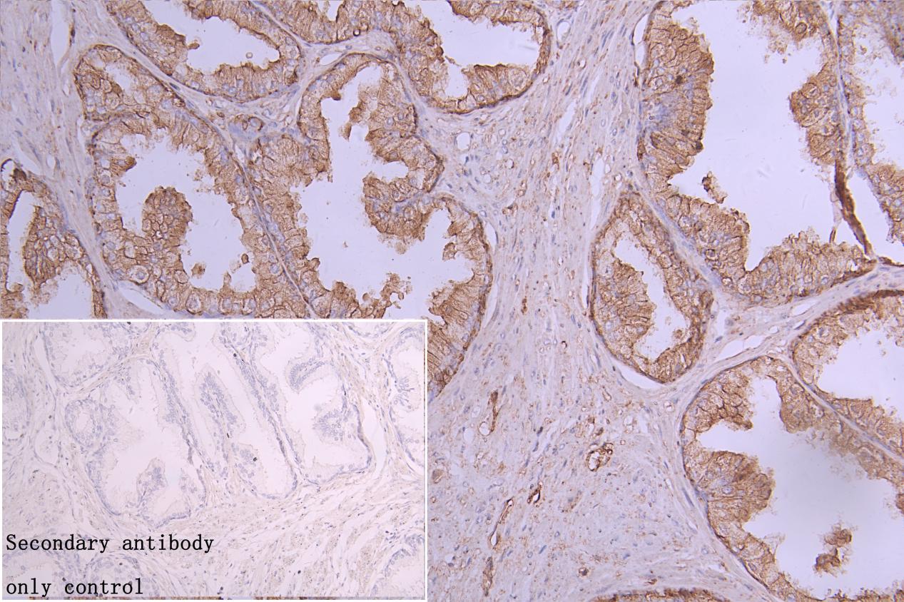
IHC image of CSB-PA10559A0Rb diluted at 1:50 and staining in paraffin-embedded human small bladder cancer performed on a Leica BondTM system. After dewaxing and hydration, antigen retrieval was mediated by high pressure in a citrate buffer (pH 6.0). Section was blocked with 10% normal goat serum 30min at RT. Then primary antibody (1% BSA) was incubated at 4C overnight. The primary is detected by a Goat anti-rabbit polymer IgG labeled by HRP and visualized using 0.05% DAB. Secondary antibody only control: uses 1% BSA instead of primary antibody |
|
|
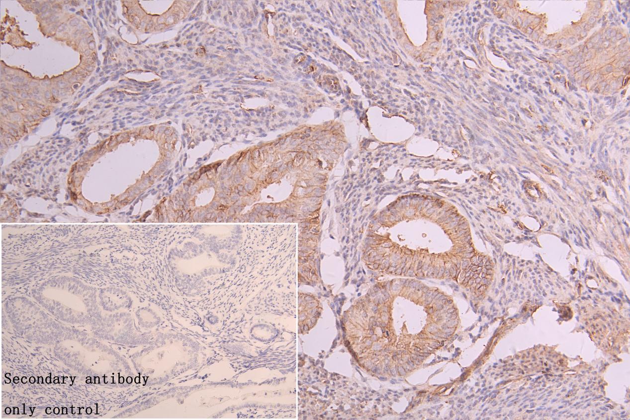
IHC image of CSB-PA10559A0Rb diluted at 1:50 and staining in paraffin-embedded human small endometrial cancer performed on a Leica BondTM system. After dewaxing and hydration, antigen retrieval was mediated by high pressure in a citrate buffer (pH 6.0). Section was blocked with 10% normal goat serum 30min at RT. Then primary antibody (1% BSA) was incubated at 4C overnight. The primary is detected by a Goat anti-rabbit polymer IgG labeled by HRP and visualized using 0.05% DAB. Secondary antibody only control: uses 1% BSA instead of primary antibody |
|
|
IHC image of CSB-PA10559A0Rb diluted at 1:50 and staining in paraffin-embedded human small prostate cancer performed on a Leica BondTM system. After dewaxing and hydration, antigen retrieval was mediated by high pressure in a citrate buffer (pH 6.0). Section was blocked with 10% normal goat serum 30min at RT. Then primary antibody (1% BSA) was incubated at 4C overnight. The primary is detected by a Goat anti-rabbit polymer IgG labeled by HRP and visualized using 0.05% DAB. Secondary antibody only control: uses 1% BSA instead of primary antibody |
|
|
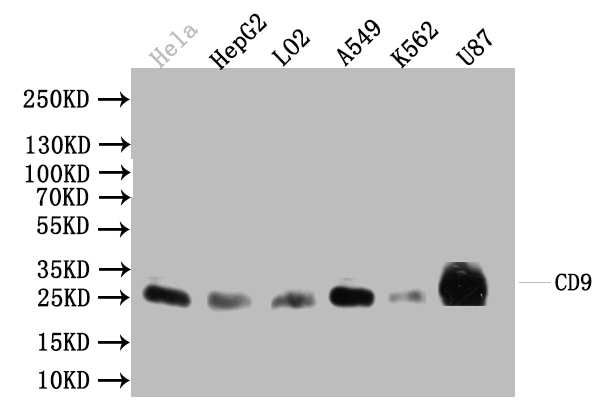
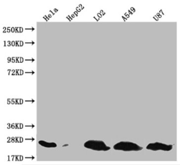
Western Blot Positive WB detected in: Hela whole cell lysate(30µg), HepG2 whole cell lysate(30µg), L02 whole cell lysate(30µg), A549 whole cell lysate(30µg), K562 whole cell lysate(30µg) All lanes: CD9 antibody at 1:1000 Secondary Goat polyclonal to rabbit IgG at 1/20000 dilution Predicted band size: 23-30 kDa Observed band size: 23 kDa Exposure time:120s |
|
|
Product Guarantee and Expert Support
