
UCP1 Antibody, Unconjugated, Rabbit, Polyclonal
Catalog Number:
CSB-PA599294GA01HU
- Images (5)
| Article Name: | UCP1 Antibody, Unconjugated, Rabbit, Polyclonal |
| Biozol Catalog Number: | CSB-PA599294GA01HU |
| Supplier Catalog Number: | CSB-PA599294GA01HU |
| Alternative Catalog Number: | CSB-PA599294GA01HU-100UL |
| Manufacturer: | Cusabio |
| Host: | Rabbit |
| Category: | Antikörper |
| Application: | ELISA, IF, IHC, WB |
| Species Reactivity: | Human, Mouse |
| Conjugation: | Unconjugated |
| Alternative Names: | mitochondrial brown fat uncoupling protein antibody, Mitochondrial brown fat uncoupling protein 1 antibody, SLC25A7 antibody, Solute carrier family 25 member 7 antibody, Thermogenin antibody, UCP 1 antibody, UCP antibody, UCP1 antibody, UCP1_HUMAN antibody, uncoupling protein 1 (mitochondrial, proton carrier) antibody, Uncoupling protein 1 antibody |
| Clonality: | Polyclonal |
| UniProt: | P25874 |
| Buffer: | PBS with 0.1% Sodium Azide, 50% Glycerol, pH 7.3. -20°C, Avoid freeze / thaw cycles. |
| Purity: | Antigen Affinity Purified |
| Form: | Liquid |
| Target: | UCP1 |
| Application Dilute: | Recommended dilution: WB:1:1000-1:5000, IHC : 1:50-1:500, IF:1:20-1:200 |
|
|
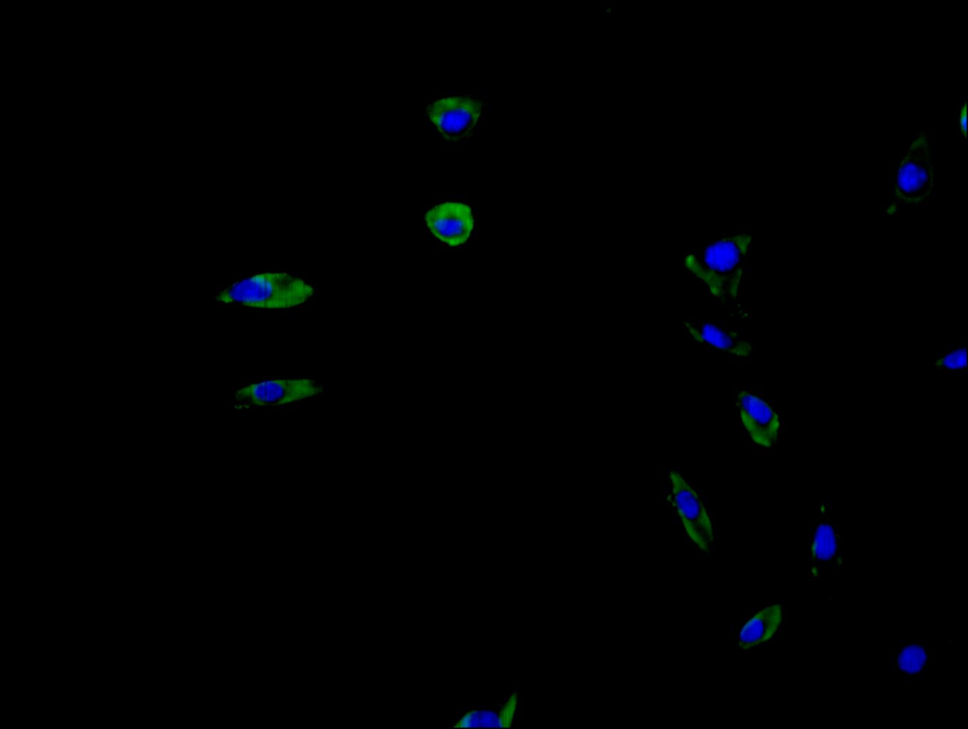
Immunofluorescence staining of SH-SY5Y cell with CSB-PA025554LA01HU at 1:20, counter-stained with DAPI. The cells were fixed in 4% formaldehyde and blocked in 10% normal Goat Serum. The cells were then incubated with the antibody overnight at 4C. The secondary antibody was Alexa Fluor 488-congugated AffiniPure Goat Anti-Rabbit IgG(H+L). |
|
|
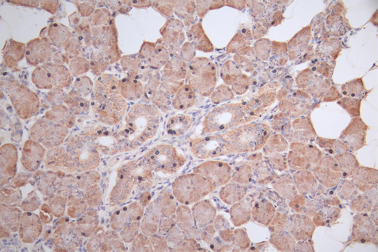
IHC image of CSB-PA025554LA01HU diluted at 1:50 and staining in paraffin-embedded human salivary gland tissue performed on a Leica BondTM system. After dewaxing and hydration, antigen retrieval was mediated by high pressure in a citrate buffer (pH 6.0). Section was blocked with 10% normal goat serum 30min at RT. Then primary antibody (1% BSA) was incubated at 4C overnight. The primary is detected by a Goat anti-rabbit polymer IgG labeled by HRP and visualized using 0.05% DAB. |
|
|
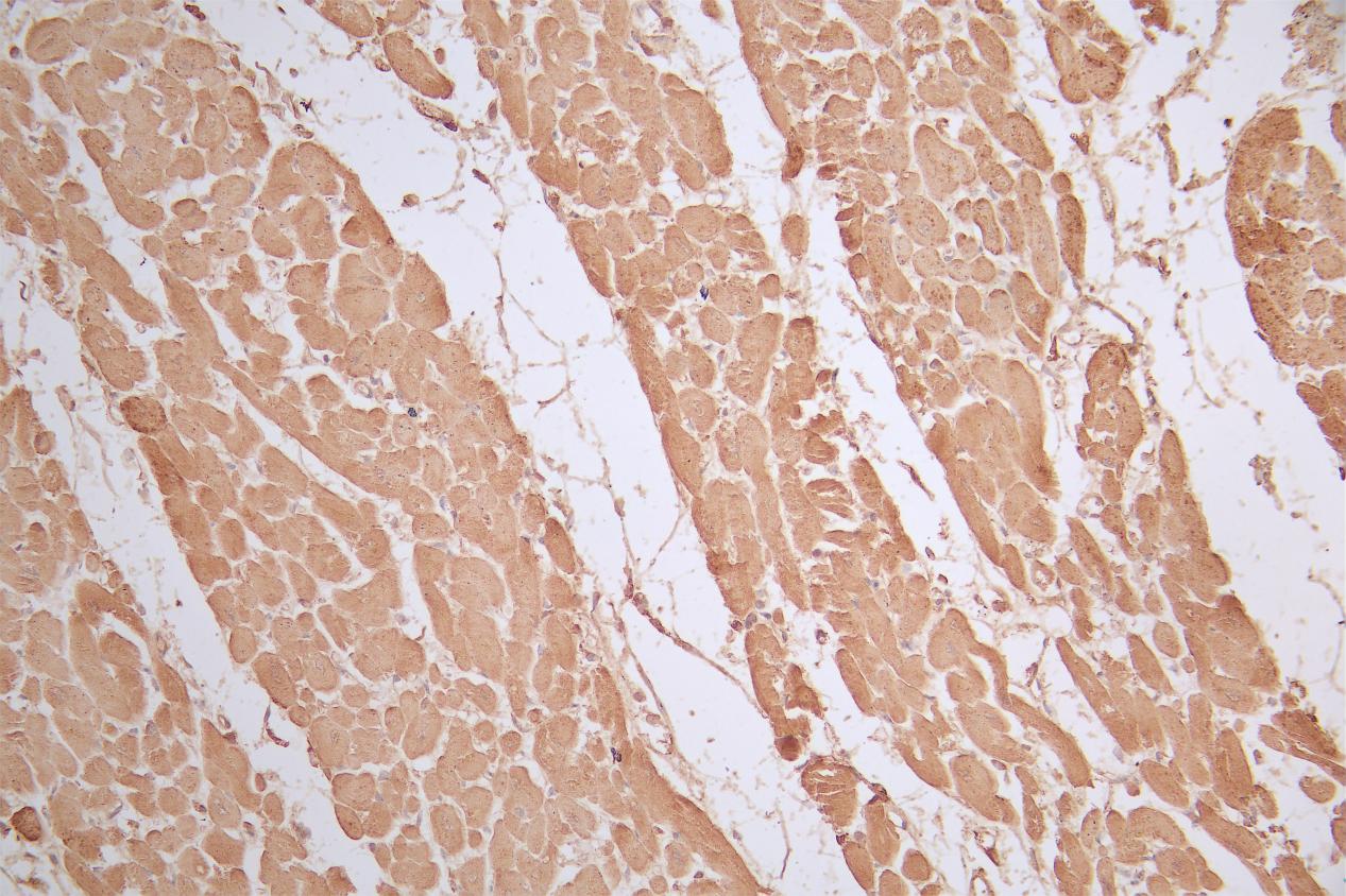
IHC image of CSB-PA025554LA01HU diluted at 1:50 and staining in paraffin-embedded human heart tissue performed on a Leica BondTM system. After dewaxing and hydration, antigen retrieval was mediated by high pressure in a citrate buffer (pH 6.0). Section was blocked with 10% normal goat serum 30min at RT. Then primary antibody (1% BSA) was incubated at 4C overnight. The primary is detected by a Goat anti-rabbit polymer IgG labeled by HRP and visualized using 0.05% DAB. |
|
|
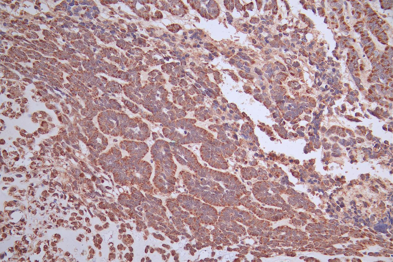
IHC image of CSB-PA025554LA01HU diluted at 1:50 and staining in paraffin-embedded human ovarian cancer performed on a Leica BondTM system. After dewaxing and hydration, antigen retrieval was mediated by high pressure in a citrate buffer (pH 6.0). Section was blocked with 10% normal goat serum 30min at RT. Then primary antibody (1% BSA) was incubated at 4C overnight. The primary is detected by a Goat anti-rabbit polymer IgG labeled by HRP and visualized using 0.05% DAB. |
|
|
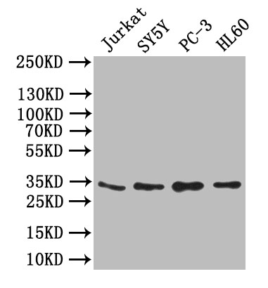
Western Blot Positive WB detected in: JK whole cell lysate,SY5Y whole cell lysate,PC-3 whole cell lysate,,HL60 whole cell lysate All lanes: UCP1 antibody at 1:1000 Secondary Goat polyclonal to rabbit IgG at 1/50000 dilution Predicted band size: 33 kDa Observed band size: 33 kDa |
Product Guarantee and Expert Support
