
MEST Antibody, Unconjugated, Rabbit, Polyclonal
Catalog Number:
CSB-PA704858ESR1HU
- Images (6)


| Article Name: | MEST Antibody, Unconjugated, Rabbit, Polyclonal |
| Biozol Catalog Number: | CSB-PA704858ESR1HU |
| Supplier Catalog Number: | CSB-PA704858ESR1HU |
| Alternative Catalog Number: | CSB-PA704858ESR1HU-100UL, CSB-PA704858ESR1HU-50UL |
| Manufacturer: | Cusabio |
| Host: | Rabbit |
| Category: | Antikörper |
| Application: | ELISA, IHC, WB |
| Species Reactivity: | Human, Mouse |
| Conjugation: | Unconjugated |
| Alternative Names: | DKFZp686L18234 antibody, EC 3.-.-.- antibody, Mesoderm specific transcript (mouse) homolog antibody, Mesoderm specific transcript homolog (mouse) antibody, Mesoderm-specific transcript homolog protein antibody, MEST antibody, MEST_HUMAN antibody, MGC111102 antibody, MGC8703 antibody, OTTHUMP00000210362 antibody, OTTHUMP00000210364 antibody, OTTHUMP00000210367 antibody, Paternally expressed gene 1 antibody, Paternally-expressed gene 1 protein antibody, PEG1 antibody |
| Clonality: | Polyclonal |
| UniProt: | Q5EB52 |
| Buffer: | PBS with 0.02% sodium azide, 50% glycerol, pH7.3. |
| Purity: | Antigen Affinity Purified |
| Form: | Liquid |
| Target: | MEST |
| Application Dilute: | Recommended dilution: WB:1:1000-1:5000, IHC:1:20-1:200 |
|
|
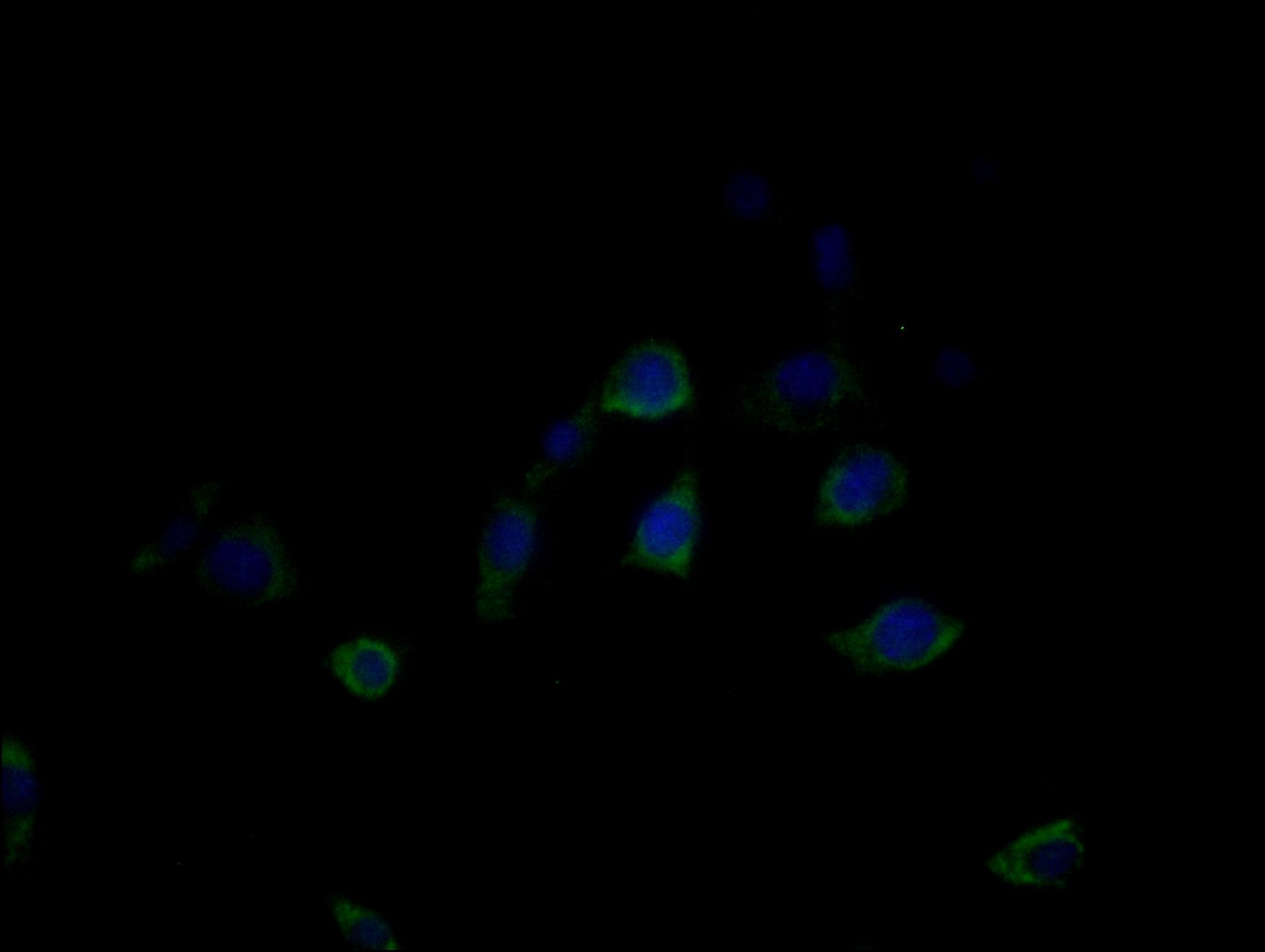
Immunofluorescence staining of MCF-7 cells with CSB-PA704858ESR1HU at 1:25, counter-stained with DAPI. The cells were fixed in 4% formaldehyde, permeabilized using 0.2% Triton X-100 and blocked in 10% normal Goat Serum. The cells were then incubated with the antibody overnight at 4C. The secondary antibody was Alexa Fluor 488-congugated AffiniPure Goat Anti-Rabbit IgG(H+L). |
|
|
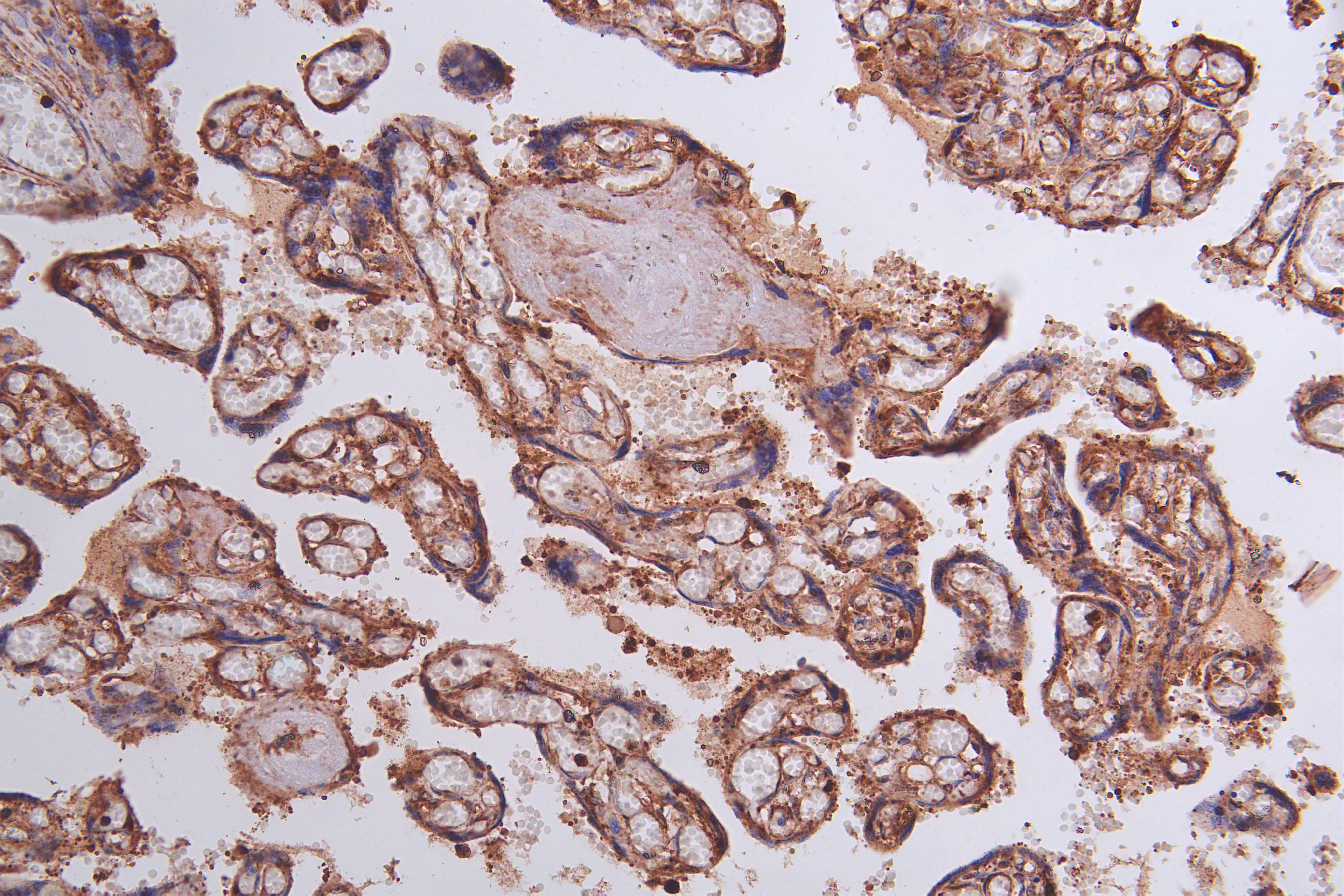
Immunohistochemistry of paraffin-embedded human pancreatic tissue using CSB-PA704858ESR1HU at dilution of 1:100 |
|
|
Immunohistochemistry of paraffin-embedded human Pancreatic tissue using CSB-PA704858ESR1HU at dilution of 1:50 |
|
|
Immunohistochemistry of paraffin-embedded human Breast cancer using CSB-PA704858ESR1HU at dilution of 1:50 |
|
|
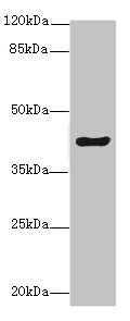
Western blot All lanes: MEST antibody at 5.78ug/ml + Mouse kidney tissue Secondary Goat polyclonal to rabbit IgG at 1/10000 dilution Predicted band size: 39, 38, 34 kDa Observed band size: 39 kDa |
|
|
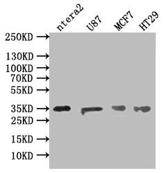
Western Blot Positive WB detected in: NTERA-2 whole cell lysate, U87 whole cell lysate, MCF7 whole cell lysate, HT29 whole cell lysate All lanes: MEST antibody at 1:500 Secondary Goat polyclonal to rabbit IgG at 1/50000 dilution Predicted band size: 39, 38, 34 kDa Observed band size: 35 kDa |
Product Guarantee and Expert Support
