

NECTIN4 Antibody, Unconjugated, Rabbit, Polyclonal
Catalog Number:
CSB-PA822274LA01HU
- Images (4)
| Article Name: | NECTIN4 Antibody, Unconjugated, Rabbit, Polyclonal |
| Biozol Catalog Number: | CSB-PA822274LA01HU |
| Supplier Catalog Number: | CSB-PA822274LA01HU |
| Alternative Catalog Number: | CSB-PA822274LA01HU-100UG, CSB-PA822274LA01HU-50UG |
| Manufacturer: | Cusabio |
| Host: | Rabbit |
| Category: | Antikörper |
| Application: | ELISA, IF |
| Species Reactivity: | Human |
| Conjugation: | Unconjugated |
| Alternative Names: | DKFZp686K05193 antibody, EDSS1 antibody, Ig superfamily receptor LNIR antibody, Nectin 4 antibody, Nectin-4 antibody, poliovirus receptor-related 4 antibody, Processed poliovirus receptor-related protein 4 antibody, PRR4 antibody, pvrl4 antibody, PVRL4_HUMAN antibody |
| Clonality: | Polyclonal |
| UniProt: | Q96NY8 |
| Buffer: | PBS with 0.02% sodium azide, 50% glycerol, pH7.3. |
| Purity: | >95%, Protein G purified |
| Form: | Liquid |
| Target: | NECTIN4 |
| Application Dilute: | Recommended dilution: IF:1:50-1:200 |
|
|
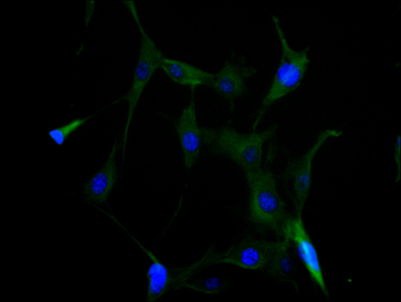
Immunofluorescence staining of NIH/3T3 cell with CSB-PA822274LA01HU at 1:35, counter-stained with DAPI. The cells were fixed in 4% formaldehyde and blocked in 10% normal Goat Serum. The cells were then incubated with the antibody overnight at 4C. The secondary antibody was Alexa Fluor 488-congugated AffiniPure Goat Anti-Rabbit IgG(H+L). |
|
|
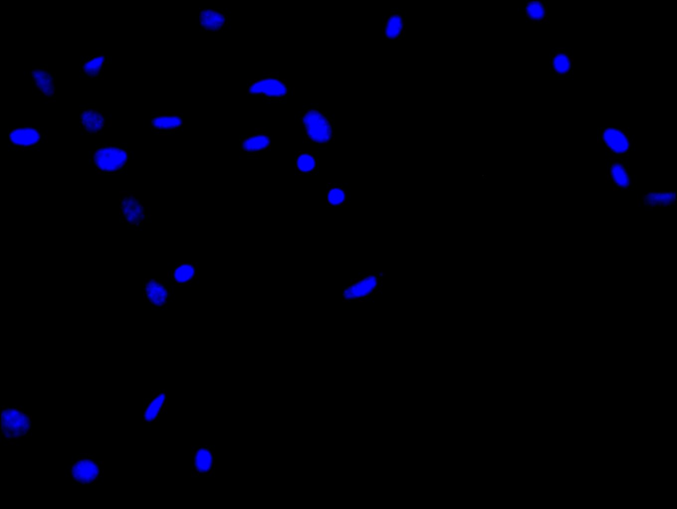
Immunofluorescence staining of NIH/3T3 cell with 5% goat serum, counter-stained with DAPI. The cells were fixed in 4% formaldehyde and blocked in 10% normal Goat Serum. The cells were then incubated with the antibody overnight at 4C. The secondary antibody was Alexa Fluor 488-congugated AffiniPure Goat Anti-Rabbit IgG(H+L). |
|
|
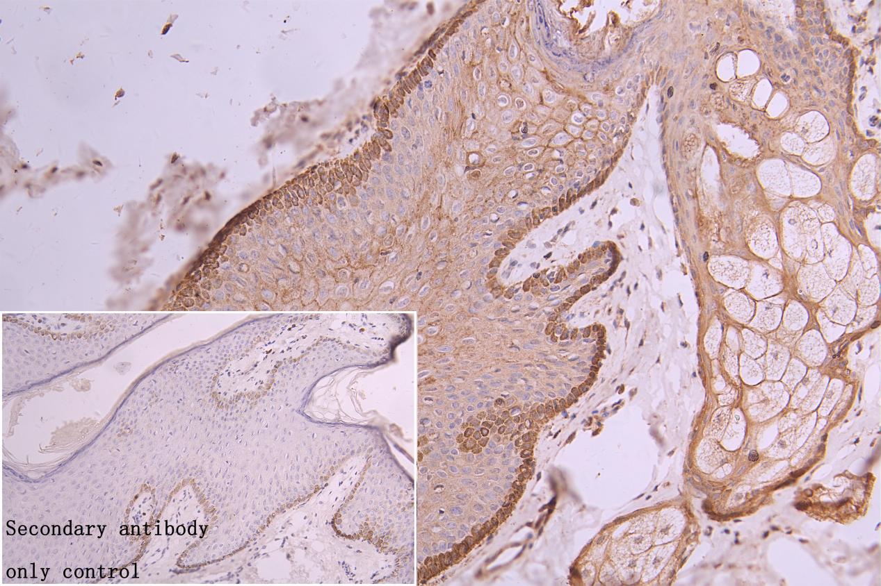
IHC image of CSB-PA822274LA01HU diluted at 1:300 and staining in paraffin-embedded human skin tissue performed on a Leica BondTM system. After dewaxing and hydration, antigen retrieval was mediated by high pressure in a citrate buffer (pH 6.0). Section was blocked with 10% normal goat serum 30min at RT. Then primary antibody (1% BSA) was incubated at 4C overnight. The primary is detected by a Goat anti-rabbit polymer IgG labeled by HRP and visualized using 0.05% DAB.Secondary antibody only control: uses 1% BSA instead of primary antibody |
|
|
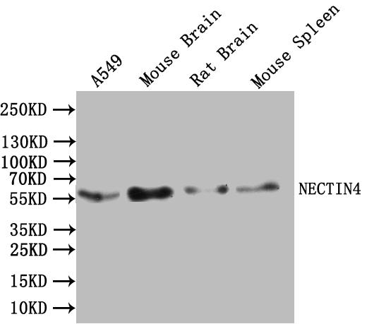
Western Blot Positive WB detected in:A549 whole cell lysate, Mouse Brain tissue lysate, Rat Brain tissue lysate, Mouse Spleen tissue lysate All lanes:NECTIN4 Antibody at 1:1000 Secondary Goat polyclonal to rabbit IgG at 1/50000 dilution Predicted band size: 56kDa Observed band size: 56 kDa |
Product Guarantee and Expert Support
