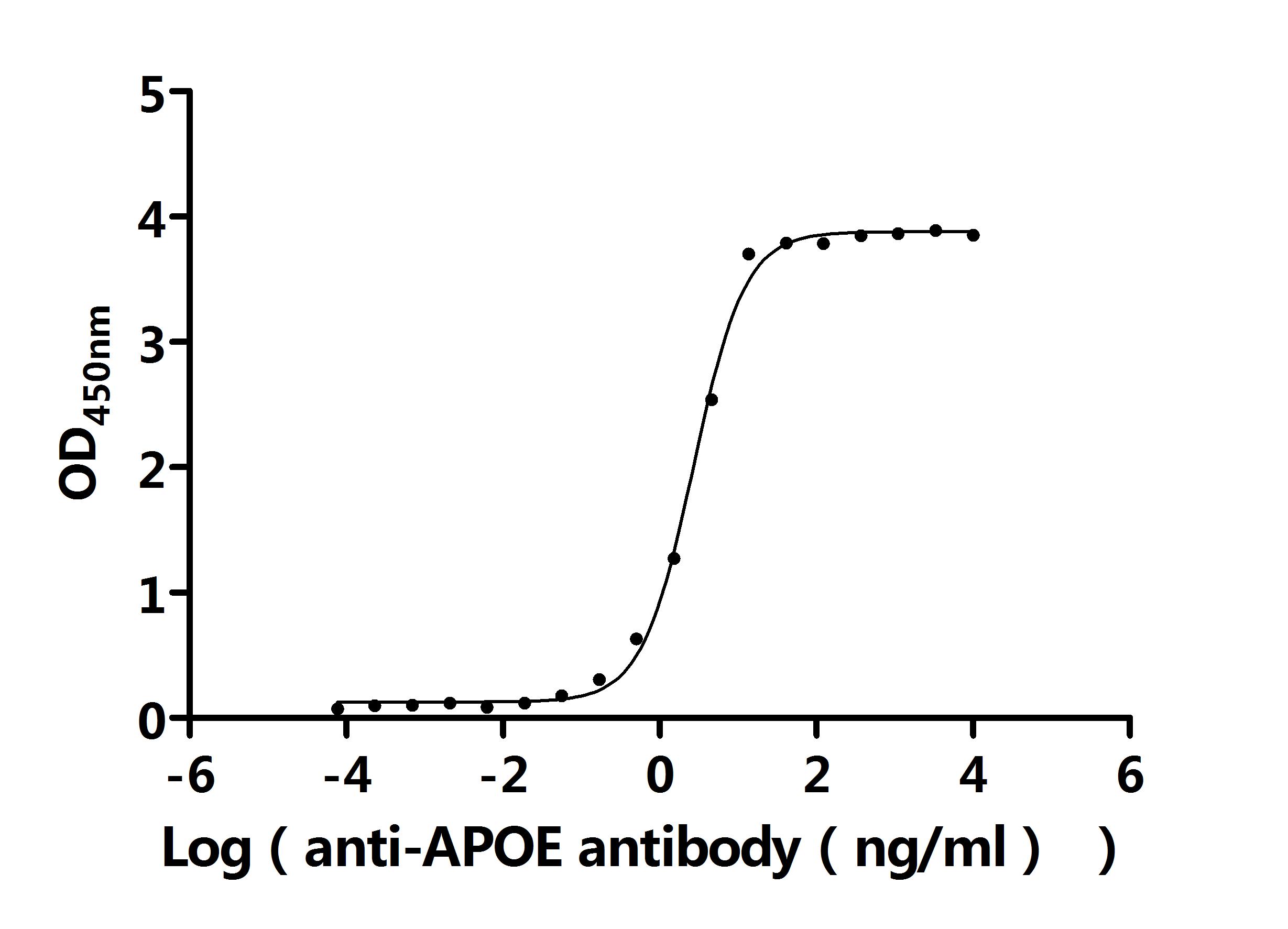
The Binding Activity of Human APOE with Anti-APOE recombinant antibody Activity: Measured by its binding ability in a functional ELISA. Immobilized Human APOE (CSB-MP001936HU) at 2 µg/mL can bind Anti-APOE recombinant antibody. The EC50 is 2.491-2.918 ng/mL.
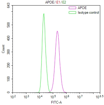
Overlay Peak curve showing HepG2 cells stained with CSB-RA001936MA1HU (red line) at 1:100. The cells were fixed in 4% formaldehyde and permeated by 0.2% TritonX-100 for10min. Then 10% normal goat serum to block non-specific protein-protein interactions followed by the antibody (1ug/1*106cells) for 45min at 4°C. The secondary antibody used was FITC-conjugatedGoatAnti-MouseIgG(H+L) at 1:200 dilution for 35min at 4°C.Control antibody (green line) was mouse IgG2a (1ug/1*106cells) used under the same conditions. Acquisition of >10,000 events was performed.
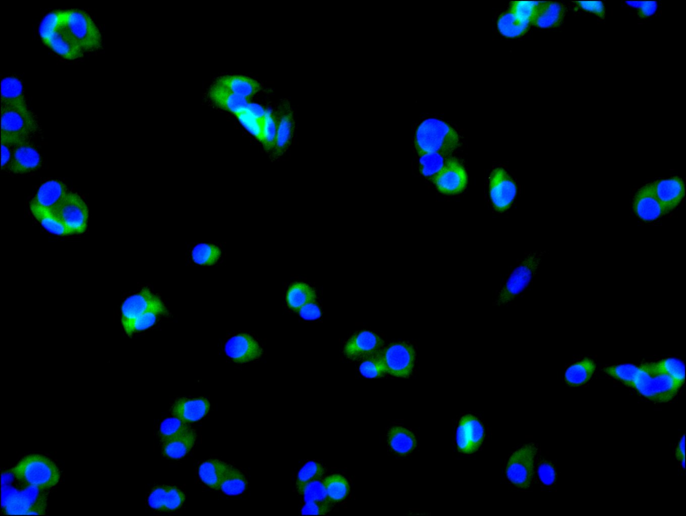
Immunofluorescence staining of HepG2 cell with CSB-RA001936MA1HU at 1:30 ,counter-stained with DAPI. The cells were fixed in 4% formaldehyde, permeabilized using 0.2% Triton X-100 and blocked in 10% normal Goat Serum. The cells were then incubated with the antibody overnight at 4C. The secondary antibody was FITC-conjugatedGoatAnti-MouseIgG(H+L).
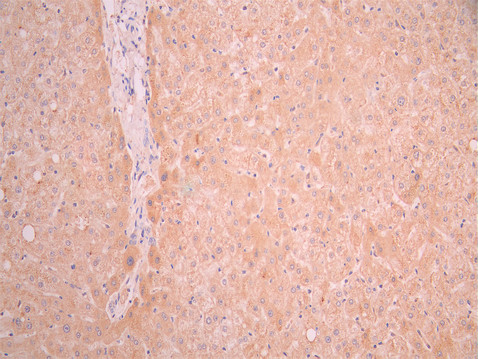
IHC image of CSB-RA001936MA1HU diluted at 1:50 and staining in paraffin-embedded human liver tissue performed on a Leica BondTM system. After dewaxing and hydration, antigen retrieval was mediated by high pressure in a citrate buffer (pH 6.0). Section was blocked with 10% normal goat serum 30min at RT. Then primary antibody (1% BSA) was incubated at 4C overnight. The primary is detected by a Goat anti-mouse polymer IgG labeled by HRP and visualized using 0.05% DAB.
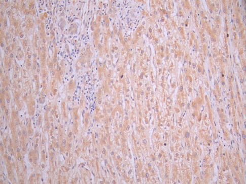
IHC image of CSB-RA001936MA1HU diluted at 1:50 and staining in paraffin-embedded human liver cancer performed on a Leica BondTM system. After dewaxing and hydration, antigen retrieval was mediated by high pressure in a citrate buffer (pH 6.0). Section was blocked with 10% normal goat serum 30min at RT. Then primary antibody (1% BSA) was incubated at 4C overnight. The primary is detected by a Goat anti-mouse polymer IgG labeled by HRP and visualized using 0.05% DAB.
* VAT and and shipping costs not included. Errors and price changes excepted