A I antibody, Al antibody, ARG 1 antibody, arg1 antibody, ARGI1_HUMAN antibody, Arginase 1 antibody, Arginase liver antibody, Arginase type I antibody, Arginase, liver antibody, Arginase-1 antibody, Arginase1 antibody, Liver type arginase antibody, Liver-type arginase antibody, Type I arginase antibody
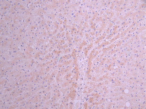
IHC image of CSB-RA002005MA1HU diluted at 1:50 and staining in paraffin-embedded human liver tissue performed on a Leica BondTM system. After dewaxing and hydration, antigen retrieval was mediated by high pressure in a citrate buffer (pH 6.0). Section was blocked with 10% normal goat serum 30min at RT. Then primary antibody (1% BSA) was incubated at 4C overnight. The primary is detected by a Anti-Human lgG, Fcy Fragment Specific labeled by HRP and visualized using 0.05% DAB.
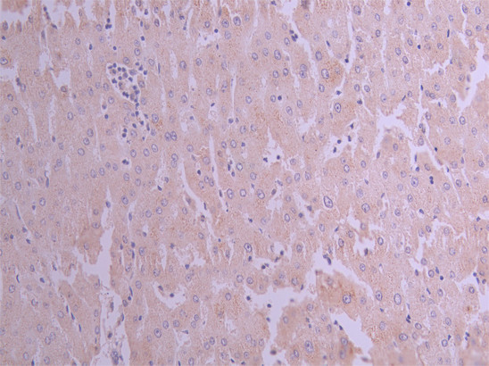
IHC image of CSB-RA002005MA1HU diluted at 1:50 and staining in paraffin-embedded human liver cancer performed on a Leica BondTM system. After dewaxing and hydration, antigen retrieval was mediated by high pressure in a citrate buffer (pH 6.0). Section was blocked with 10% normal goat serum 30min at RT. Then primary antibody (1% BSA) was incubated at 4C overnight. The primary is detected by a Anti-Human lgG, Fcy Fragment Specific labeled by HRP and visualized using 0.05% DAB.
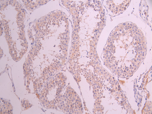
IHC image of CSB-RA002005MA1HU diluted at 1:50 and staining in paraffin-embedded human testis tissue performed on a Leica BondTM system. After dewaxing and hydration, antigen retrieval was mediated by high pressure in a citrate buffer (pH 6.0). Section was blocked with 10% normal goat serum 30min at RT. Then primary antibody (1% BSA) was incubated at 4C overnight. The primary is detected by a Anti-Human lgG, Fcy Fragment Specific labeled by HRP and visualized using 0.05% DAB.
* VAT and and shipping costs not included. Errors and price changes excepted