
CYP19A1 Recombinant Monoclonal Antibody, Clone: [1H1], Unconjugated, Rabbit
Catalog Number:
CSB-RA006394A0HU
- Images (5)
| Article Name: | CYP19A1 Recombinant Monoclonal Antibody, Clone: [1H1], Unconjugated, Rabbit |
| Biozol Catalog Number: | CSB-RA006394A0HU |
| Supplier Catalog Number: | CSB-RA006394A0HU |
| Alternative Catalog Number: | CSB-RA006394A0HU-100UL, CSB-RA006394A0HU-50UL |
| Manufacturer: | Cusabio |
| Host: | Rabbit |
| Category: | Antikörper |
| Application: | ELISA, IF, IHC, WB |
| Species Reactivity: | Human |
| Conjugation: | Unconjugated |
| Alternative Names: | Aromatase, CYPXIX, Cytochrome P-450AROM, Cytochrome P450 19A1, Estrogen synthase, CYP19A1, ARO1, CYAR, CYP19 |
| Clonality: | Monoclonal |
| Clone Designation: | [1H1] |
| UniProt: | P11511 |
| Buffer: | Rabbit IgG in 10mM phosphate buffered saline , pH 7.4, 150mM sodium chloride, 0.05% BSA, 0.02% sodium azide and 50% glycerol. |
| Purity: | Affinity-chromatography |
| Form: | Liquid |
| Target: | CYP19A1 |
| Antibody Type: | Recombinant Antibody |
| Application Dilute: | Recommended dilution: WB:1:500-1:5000, IHC:1:200-1:500, IF:1:20-1:200 |
|
|
|
|
|
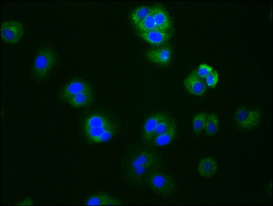
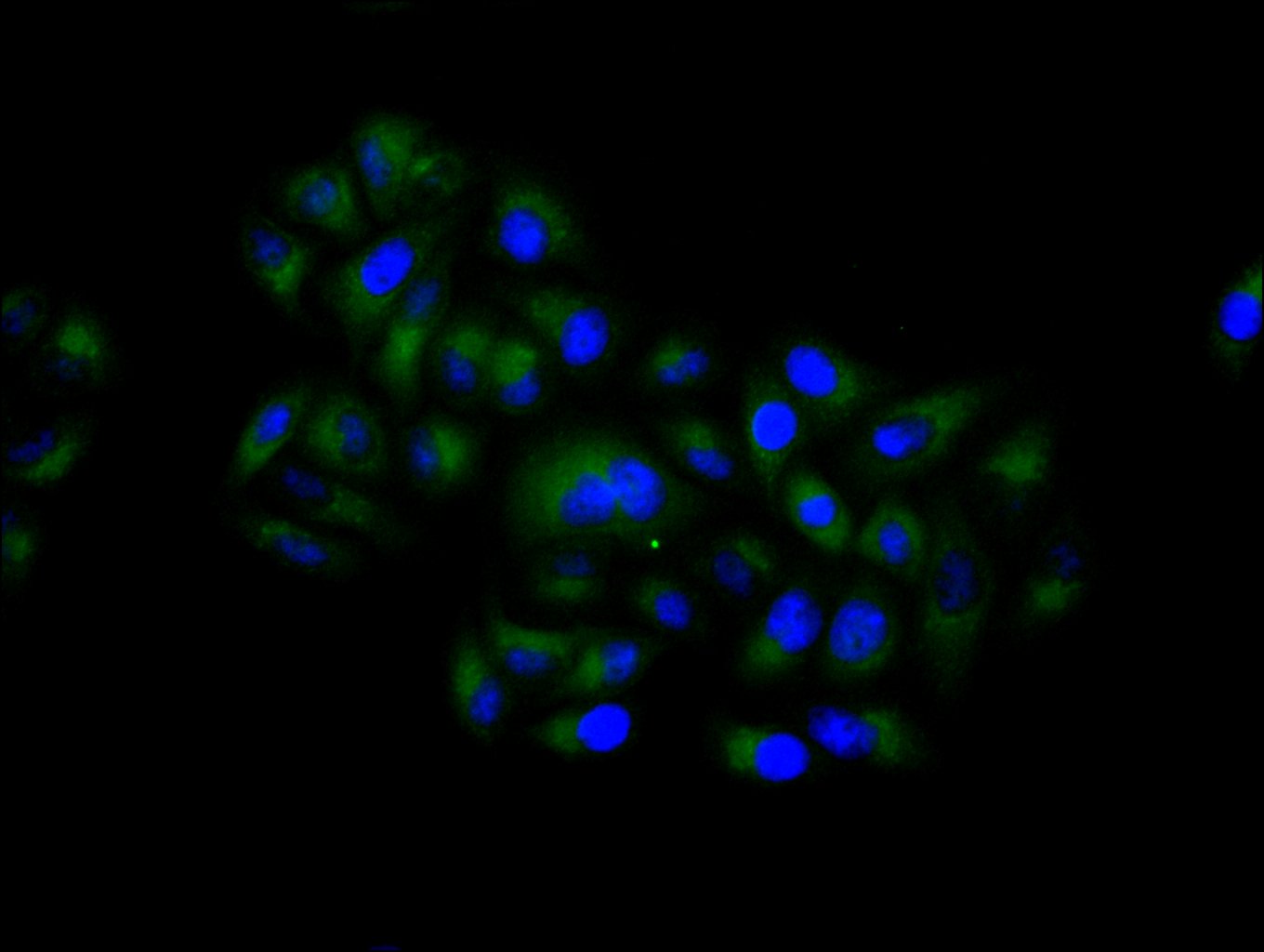
Immunofluorescence staining of Hela cell with CSB-RA006394A0HU at 1:50, counter-stained with DAPI. The cells were fixed in 4% formaldehyde and blocked in 10% normal Goat Serum. The cells were then incubated with the antibody overnight at 4C. The secondary antibody was Alexa Fluor 488-congugated AffiniPure Goat Anti-Rabbit IgG(H+L) . |
|
|
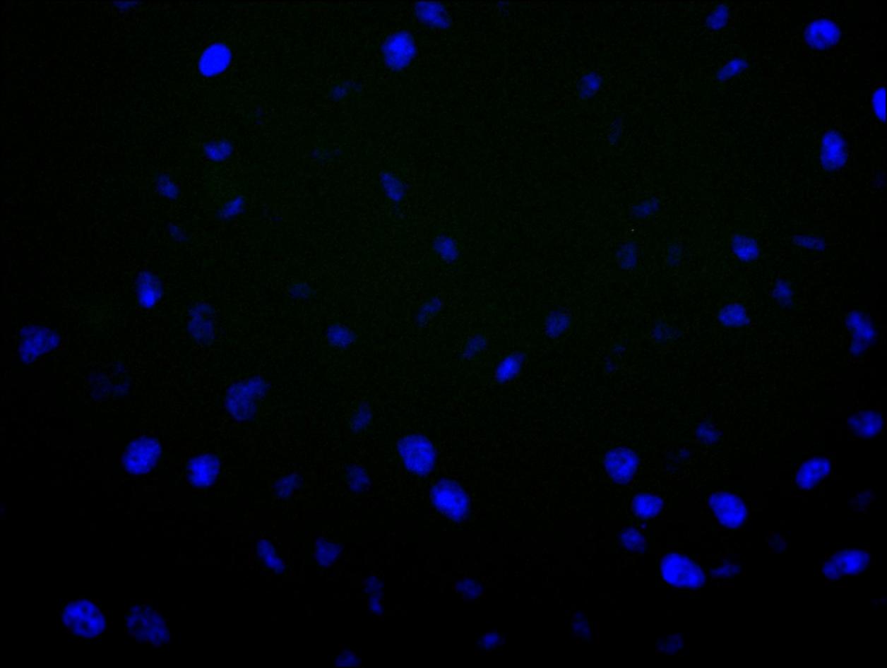
Immunofluorescence staining of Hela cell with 5% goat serum, counter-stained with DAPI. The cells were fixed in 4% formaldehyde and blocked in 10% normal Goat Serum. The cells were then incubated with the antibody overnight at 4C. The secondary antibody was Alexa Fluor 488-congugated AffiniPure Goat Anti-Rabbit IgG(H+L) . |
|
|
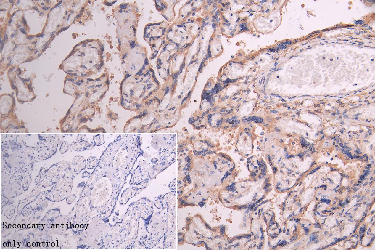
IHC image of CSB-RA006394A0HU diluted at 1:300 and staining in paraffin-embedded human placenta tissue performed on a Leica BondTM system. After dewaxing and hydration, antigen retrieval was mediated by high pressure in a citrate buffer (pH 6.0) . Section was blocked with 10% normal goat serum 30min at RT. Then primary antibody (1% BSA) was incubated at 4C overnight. The primary is detected by a Goat anti-rabbit polymer IgG labeled by HRP and visualized using 0.05% DAB.Secondary antibody only control: uses 1% BSA instead of primary antibody |
|
|
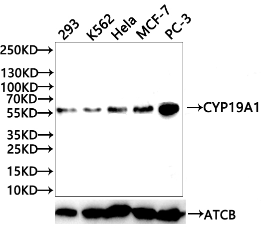
Western Blot Positive WB detected in: 293 whole cell lysate(20µg) , K562 whole cell lysate(20µg) , Hela whole cell lysate(20µg) , MCF-7 whole cell lysate(20µg) , PC-3 whole cell lysate(20µg) All lanes: CYP19A1 antibody at 1:1000 Secondary Goat polyclonal to rabbit IgG at 1/50000 dilution Predicted band size: 58 kDa Observed band size: 58 kDa Exposure time:120s |
Product Guarantee and Expert Support
