
FLOT1 Recombinant Monoclonal Antibody, Clone: [4C8B6], Unconjugated
Catalog Number:
CSB-RA008727MA1HU
- Images (5)
| Article Name: | FLOT1 Recombinant Monoclonal Antibody, Clone: [4C8B6], Unconjugated |
| Biozol Catalog Number: | CSB-RA008727MA1HU |
| Supplier Catalog Number: | CSB-RA008727MA1HU |
| Alternative Catalog Number: | CSB-RA008727MA1HU-100UL, CSB-RA008727MA1HU-50UL |
| Manufacturer: | Cusabio |
| Category: | Antikörper |
| Application: | ELISA, FC, IF, WB |
| Species Reactivity: | Human, Mouse, Rat |
| Conjugation: | Unconjugated |
| Alternative Names: | FLOT 1 antibody, FLOT1 antibody, FLOT1_HUMAN antibody, Flotillin-1 antibody, Flotillin1 antibody, Integral membrane component of caveolae antibody, Reggie 2 antibody |
| Clonality: | Monoclonal |
| Clone Designation: | [4C8B6] |
| UniProt: | O75955 |
| Buffer: | Preservative: 0.03% Proclin 300<br />Constituents: 50% Glycerol, 0.01M PBS, PH 7.4 |
| Purity: | Affinity-chromatography |
| Form: | Liquid |
| Target: | FLOT1 |
| Antibody Type: | Recombinant Antibody |
| Application Dilute: | Recommended dilution: WB:1:500-1:2000, IF:1:50-1:200, FC:1:50-1:200 |
|
|
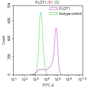
Overlay Peak curve showing A431 cells stained with CSB-RA008727MA1HU (red line) at 1:100. The cells were fixed in 4% formaldehyde and permeated by 0.2% TritonX-100 for10min. Then 10% normal goat serum to block non-specific protein-protein interactions followed by the antibody (1ug/1*106cells) for 45min at 4°C. The secondary antibody used was Fluorescein (FITC) AffiniPure Goat Anti-Human IgG, Fcgamma fragment specific at 1:200 dilution for 35min at 4°C.Control antibody (green line) was human IgG1 (1ug/1*106cells) used under the same conditions. Acquisition of >10,000 events was performed. |
|
|
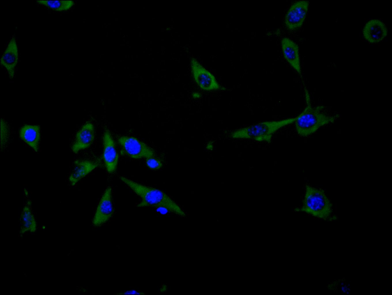
Immunofluorescence staining of NIH/3T3 cell with CSB-RA008727MA1HU at 1:30 counter-stained with DAPI. The cells were fixed in 4% formaldehyde and blocked in 10% normal Goat Serum. The cells were then incubated with the antibody overnight at 4C. The secondary antibody was Fluorescein (FITC) AffiniPure Goat Anti-Human IgG, Fcgamma fragment specific. |
|
|
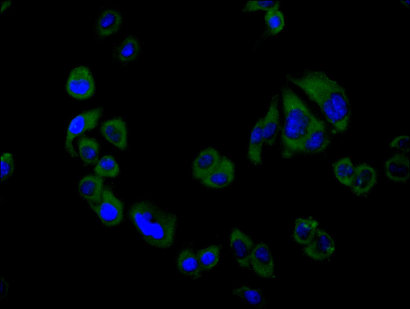
Immunofluorescence staining of Hela cell with CSB-RA008727MA1HU at 1:30 counter-stained with DAPI. The cells were fixed in 4% formaldehyde and blocked in 10% normal Goat Serum. The cells were then incubated with the antibody overnight at 4C. The secondary antibody was Fluorescein (FITC) AffiniPure Goat Anti-Human IgG, Fcgamma fragment specific. |
|
|
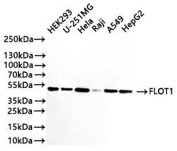
Western Blot Positive WB detected in: HEK293 whole cell lysate(20µg) , U-251MG whole cell lysate(20µg) , HeLa whole cell lysate(20µg) , Raji whole cell lysate(20µg) , A549 whole cell lysate(20µg) , HepG2 whole cell lysate(20µg) All lanes: FLOT1 antibody at 1:1000 Secondary Goat polyclonal to human IgG at 1/40000 dilution Predicted band size: 47 kDa Observed band size: 47 kDa Exposure time:1min |
|
|
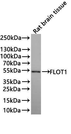
Western Blot Positive WB detected in: Rat brain tissue lysate(20µg) All lanes: FLOT1 antibody at 1:1000 Secondary Goat polyclonal to human IgG at 1/40000 dilution Predicted band size: 47.4 kDa Observed band size: 48 kDa Exposure time: 2min |
Product Guarantee and Expert Support
