
GSTM3 Recombinant Monoclonal Antibody, Clone: [2H9D7], Unconjugated
Catalog Number:
CSB-RA009982MA1HU
- Images (5)

| Article Name: | GSTM3 Recombinant Monoclonal Antibody, Clone: [2H9D7], Unconjugated |
| Biozol Catalog Number: | CSB-RA009982MA1HU |
| Supplier Catalog Number: | CSB-RA009982MA1HU |
| Alternative Catalog Number: | CSB-RA009982MA1HU-100UL, CSB-RA009982MA1HU-50UL |
| Manufacturer: | Cusabio |
| Category: | Antikörper |
| Application: | ELISA, FC, IHC, WB |
| Species Reactivity: | Human, Mouse, Rat |
| Conjugation: | Unconjugated |
| Alternative Names: | brain GST antibody, brain type mu glutathione S transferase antibody, glutathione S alkyltransferase M3 antibody, glutathione S aryltransferase M3 antibody, glutathione S transferase M3 (brain) antibody, glutathione S transferase M3 antibody, Glutathione S transferase Mu 3 antibody, glutathione S transferase, Mu 3 antibody, Glutathione S-transferase Mu 3 antibody, GST class mu 3 antibody, GST class-mu 3 antibody, GST5 antibody, GSTB antibody, Gstm3 antibody, GSTM3-3 antibody, GSTM3_HUMAN antibody, GTM3 antibody, hGSTM3-3 antibody, MGC3310 antibody, MGC3704 antibody, S (hydroxyalkyl) glutathione lyase M3 antibody |
| Clonality: | Monoclonal |
| Clone Designation: | [2H9D7] |
| UniProt: | P21266 |
| Buffer: | Preservative: 0.03% Proclin 300<br />Constituents: 50% Glycerol, 0.01M PBS, PH 7.4 |
| Purity: | Affinity-chromatography |
| Form: | Liquid |
| Target: | GSTM3 |
| Antibody Type: | Recombinant Antibody |
| Application Dilute: | Recommended dilution: WB:1:500-1:2000, IHC:1:50-1:200, FC:1:50-1:200 |
|
|
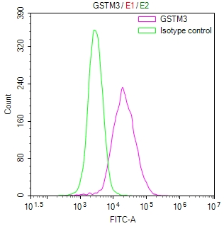
Overlay Peak curve showing MCF-7 cells stained with CSB-RA009982MA1HU (red line) at 1:100. The cells were fixed in 4% formaldehyde and permeated by 0.2% TritonX-100 for10min. Then 10% normal goat serum to block non-specific protein-protein interactions followed by the antibody (1ug/1*106cells) for 45min at 4°C. The secondary antibody used was Fluorescein (FITC) AffiniPure Goat Anti-Human IgG, Fcgamma fragment specific at 1:200 dilution for 35min at 4°C.Control antibody (green line) was human IgG1 (1ug/1*106cells) used under the same conditions. Acquisition of >10,000 events was performed. |
|
|
IHC image of CSB-RA009982MA1HU diluted at 1:100 and staining in paraffin-embedded human testis tissue performed on a Leica BondTM system. After dewaxing and hydration, antigen retrieval was mediated by high pressure in a citrate buffer (pH 6.0) . Section was blocked with 10% normal goat serum 30min at RT. Then primary antibody (1% BSA) was incubated at 4C overnight. The primary is detected by a Goat anti-human polymer IgG labeled by HRP and visualized using 0.05% DAB. |
|
|
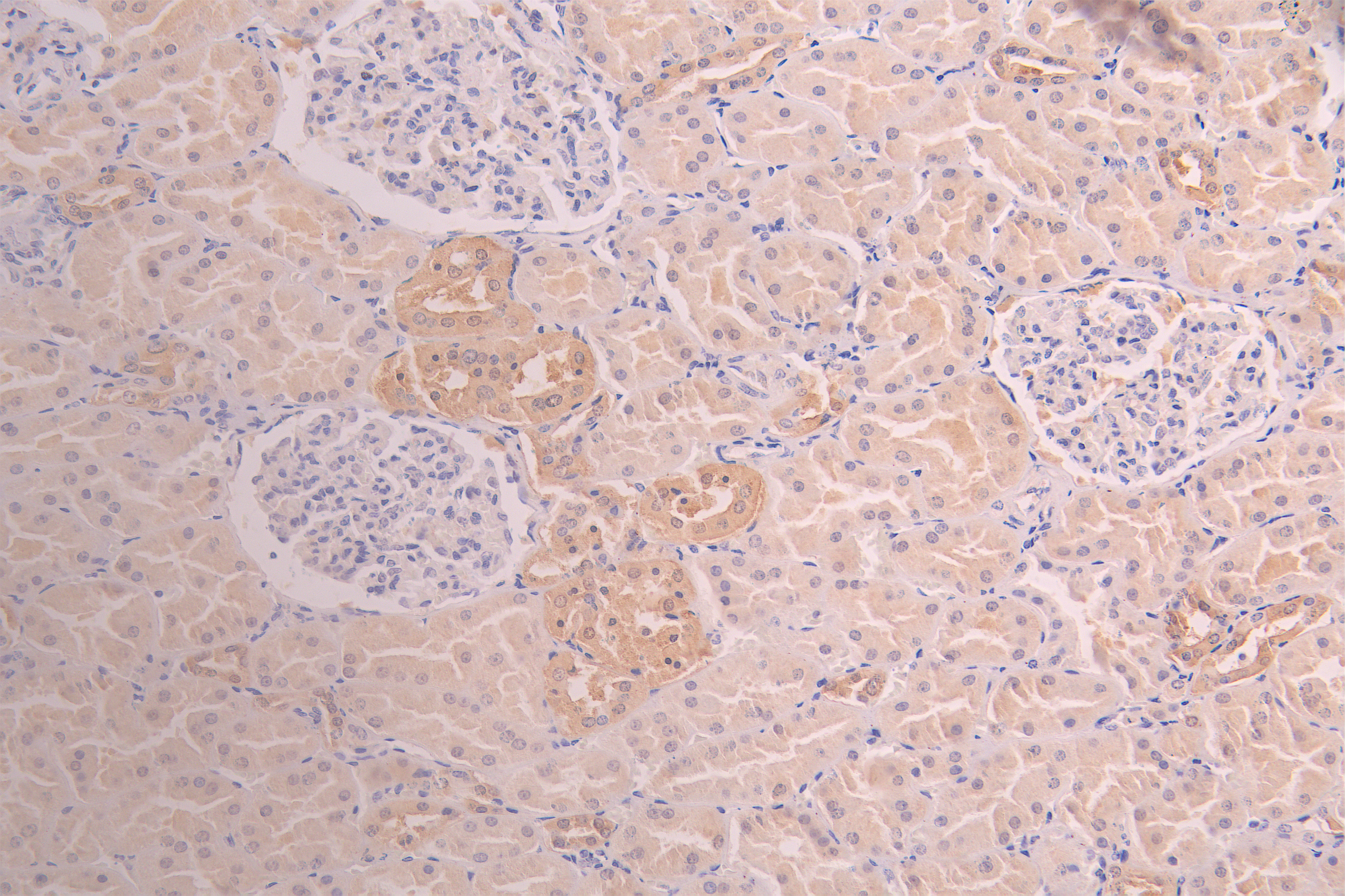
IHC image of CSB-RA009982MA1HU diluted at 1:100 and staining in paraffin-embedded human kidney tissue performed on a Leica BondTM system. After dewaxing and hydration, antigen retrieval was mediated by high pressure in a citrate buffer (pH 6.0) . Section was blocked with 10% normal goat serum 30min at RT. Then primary antibody (1% BSA) was incubated at 4C overnight. The primary is detected by a Goat anti-human polymer IgG labeled by HRP and visualized using 0.05% DAB. |
|
|
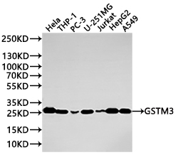
Western Blot Positive WB detected in: HeLa whole cell lysate(20µg) , THP-1 whole cell lysate(20µg) , PC-3 whole cell lysate(20µg) , U-251MG whole cell lysate(20µg) , Jurkat whole cell lysate(20µg) , HepG2 whole cell lysate(20µg) , A549 whole cell lysate(20µg) All lanes: GSTM3 antibody at 1:1000 Secondary Goat polyclonal to rabbit IgG at 1/40000 dilution Predicted band size: 27 kDa Observed band size: 27 kDa Exposure time:2min |
|
|
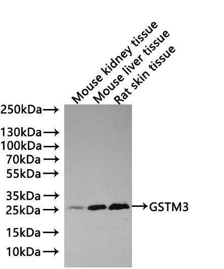
Western Blot Positive WB detected in: Mouse kidney tissue lysate(20µg) , Mouse liver tissue lysate(20µg) , Rat skin tissue lysate(20µg) All lanes: GSTM3 antibody at 1:1000 Secondary Goat polyclonal to human IgG at 1/40000 dilution Predicted band size: 26.6 kDa Observed band size: 27 kDa Exposure time: 2min |
Product Guarantee and Expert Support
