
Histone H3.1 Recombinant Monoclonal Antibody, Clone: [27F2], Unconjugated, Rabbit
Catalog Number:
CSB-RA010418A0HU
- Images (5)
| Article Name: | Histone H3.1 Recombinant Monoclonal Antibody, Clone: [27F2], Unconjugated, Rabbit |
| Biozol Catalog Number: | CSB-RA010418A0HU |
| Supplier Catalog Number: | CSB-RA010418A0HU |
| Alternative Catalog Number: | CSB-RA010418A0HU-100UL, CSB-RA010418A0HU-50UL |
| Manufacturer: | Cusabio |
| Host: | Rabbit |
| Category: | Antikörper |
| Application: | ELISA, FC, IF, IHC, WB |
| Species Reactivity: | Human |
| Conjugation: | Unconjugated |
| Alternative Names: | Histone H3.1, Histone H3/a, Histone H3/b, Histone H3/c, Histone H3/d, Histone H3/f, Histone H3/h, Histone H3/i, Histone H3/j, Histone H3/k, Histone H3/l, HIST1H3A, H3FA, AND, HIST1H3B, H3FL, AND, HIST1H3C, H3FC, AND, HIST1H3D, H3FB, AND, HIST1H3E, H3FD, AND, HIST1H3F, H3FI, AND, HIST1H3G, H3FH, AND, HIST1H3H, H3FK, AND, HIST1H3I, H3FF, AND, HIST1H3J, H3FJ |
| Clonality: | Monoclonal |
| Clone Designation: | [27F2] |
| UniProt: | P68431 |
| Buffer: | Rabbit IgG in 10mM phosphate buffered saline , pH 7.4, 150mM sodium chloride, 0.05% BSA, 0.02% sodium azide and 50% glycerol. |
| Purity: | Affinity-chromatography |
| Form: | Liquid |
| Target: | HIST1H3A |
| Antibody Type: | Recombinant Antibody |
| Application Dilute: | Recommended dilution: WB:1:500-1:5000, IHC:1:50-1:500, IF:1:30-1:200 |
|
|
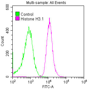
Overlay histogram showing Hela cells stained with CSB-RA010418A0HU (red line) at 1:50. The cells were fixed with 70% Ethylalcohol (18h) and then permeabilized with 0.3% Triton X-100 for 2 min.The cells were then incubated in 1x PBS /10% normal goat serum to block non-specific protein-protein interactions followed by primary antibody for 1 h at 4°C.The secondary antibody used was FITC goat anti-rabbit IgG (H+L) at 1/200 dilution for 1 h at 4°C. Control antibody (green line) was used under the same conditions. Acquisition of >10,000 events was performed. |
|
|
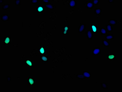
Immunofluorescence staining of Hela cells with CSB-RA010418A0HU at 1:93,counter-stained with DAPI. The cells were fixed in 4% formaldehyde, permeabilized using 0.2% Triton X-100 and blocked in 10% normal Goat Serum. The cells were then incubated with the antibody overnight at 4°C.The secondary antibody was Alexa Fluor 488-congugated AffiniPure Goat Anti-Rabbit IgG (H+L) . |
|
|
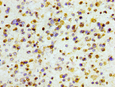
IHC image of CSB-RA010418A0HU diluted at 1:100 and staining in paraffin-embedded human glioma cancer performed on a Leica BondTM system. After dewaxing and hydration, antigen retrieval was mediated by high pressure in a citrate buffer (pH 6.0) . Section was blocked with 10% normal goat serum 30min at RT. Then primary antibody (1% BSA) was incubated at 4°C overnight. The primary is detected by a biotinylated secondary antibody and visualized using an HRP conjugated SP system. |
|
|
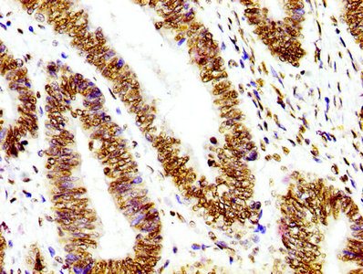
IHC image of CSB-RA010418A0HU diluted at 1:100 and staining in paraffin-embedded human colon cancer performed on a Leica BondTM system. After dewaxing and hydration, antigen retrieval was mediated by high pressure in a citrate buffer (pH 6.0) . Section was blocked with 10% normal goat serum 30min at RT. Then primary antibody (1% BSA) was incubated at 4°C overnight. The primary is detected by a biotinylated secondary antibody and visualized using an HRP conjugated SP system. |
|
|
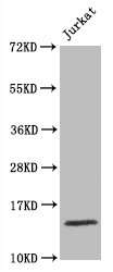
Western Blot Positive WB detected in Jurkat whole cell lysate All lanes Histone H3.1 antibody at 1.5µg/ml Secondary Goat polyclonal to rabbit IgG at 1/50000 dilution Predicted band size: 15 KDa Observed band size: 15 KDa |
Product Guarantee and Expert Support
