
HSPA5 Recombinant Monoclonal Antibody, Clone: [9G10], Unconjugated
Catalog Number:
CSB-RA010827MA1HU
- Images (5)
| Article Name: | HSPA5 Recombinant Monoclonal Antibody, Clone: [9G10], Unconjugated |
| Biozol Catalog Number: | CSB-RA010827MA1HU |
| Supplier Catalog Number: | CSB-RA010827MA1HU |
| Alternative Catalog Number: | CSB-RA010827MA1HU-100UL, CSB-RA010827MA1HU-50UL |
| Manufacturer: | Cusabio |
| Category: | Antikörper |
| Application: | ELISA, FC, IHC, WB |
| Species Reactivity: | Human |
| Conjugation: | Unconjugated |
| Alternative Names: | Endoplasmic reticulum chaperone BiP (EC 3.6.4.10) (78 kDa glucose-regulated protein) (GRP-78) (Binding-immunoglobulin protein) (BiP) (Heat shock protein 70 family protein 5) (HSP70 family protein 5) (Heat shock protein family A member 5) (Immunoglobulin heavy chain-binding protein) , HSPA5, GRP78 |
| Clonality: | Monoclonal |
| Clone Designation: | [9G10] |
| UniProt: | P11021 |
| Buffer: | Preservative: 0.03% Proclin 300<br />Constituents: 50% Glycerol, 0.01M PBS, PH 7.4 |
| Purity: | Affinity-chromatography |
| Form: | Liquid |
| Target: | HSPA5 |
| Antibody Type: | Recombinant Antibody |
| Application Dilute: | Recommended dilution: WB:1:500-1:2000, IHC:1:50-1:200, FC:1:50-1:200 |
|
|
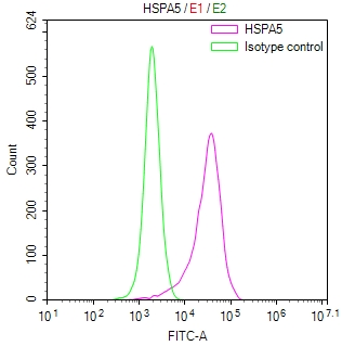
Overlay Peak curve showing U251 cells stained with CSB-RA010827MA1HU (red line) at 1:100. The cells were fixed in 4% formaldehyde and permeated by 0.2% TritonX-100 for10min. Then 10% normal goat serum to block non-specific protein-protein interactions followed by the antibody (1ug/1*106cells) for 45min at 4°C. The secondary antibody used was Fluorescein (FITC) AffiniPure Goat Anti-Human IgG, Fcgamma fragment specific at 1:200 dilution for 35 min at 4°C.Control antibody (green line) was human IgG1 (1ug/1*106cells) used under the same conditions. Acquisition of >10,000 events was performed. |
|
|
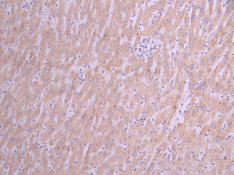
IHC image of CSB-RA010827MA1HU diluted at 1:50 and staining in paraffin-embedded human liver cancer performed on a Leica BondTM system. After dewaxing and hydration, antigen retrieval was mediated by high pressure in a citrate buffer (pH 6.0) . Section was blocked with 10% normal goat serum 30min at RT. Then primary antibody (1% BSA) was incubated at 4C overnight. The primary is detected by a Anti-Human lgG, Fcy Fragment Specific labeled by HRP and visualized using 0.05% DAB. |
|
|
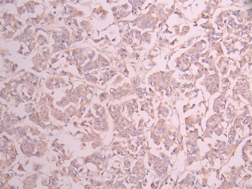
IHC image of CSB-RA010827MA1HU diluted at 1:50 and staining in paraffin-embedded human breast cancer performed on a Leica BondTM system. After dewaxing and hydration, antigen retrieval was mediated by high pressure in a citrate buffer (pH 6.0) . Section was blocked with 10% normal goat serum 30min at RT. Then primary antibody (1% BSA) was incubated at 4C overnight. The primary is detected by a Anti-Human lgG, Fcy Fragment Specific labeled by HRP and visualized using 0.05% DAB. |
|
|
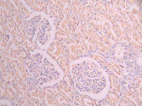
IHC image of CSB-RA010827MA1HU diluted at 1:50 and staining in paraffin-embedded human kidney tissue performed on a Leica BondTM system. After dewaxing and hydration, antigen retrieval was mediated by high pressure in a citrate buffer (pH 6.0) . Section was blocked with 10% normal goat serum 30min at RT. Then primary antibody (1% BSA) was incubated at 4C overnight. The primary is detected by a Anti-Human lgG, Fcy Fragment Specific labeled by HRP and visualized using 0.05% DAB. |
|
|
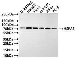
Western Blot Positive WB detected in: U-251MG whole cell lysate(20µg) , HepG2 whole cell lysate(20µg) , Hela whole cell lysate(20µg) , colo205 whole cell lysate(20µg) , A549 whole cell lysate(20µg) , PC-3 whole cell lysate(20µg) All lanes: HSPA5 antibody at 1:1000 Secondary Goat polyclonal to human IgG at 1/40000 dilution Predicted band size: 72 kDa Observed band size: 72 kDa Exposure time: 2min |
Product Guarantee and Expert Support
