
HSPA8 Recombinant Monoclonal Antibody, Clone: [2G8F6], Unconjugated
Catalog Number:
CSB-RA010829MA1HU
- Images (5)
| Article Name: | HSPA8 Recombinant Monoclonal Antibody, Clone: [2G8F6], Unconjugated |
| Biozol Catalog Number: | CSB-RA010829MA1HU |
| Supplier Catalog Number: | CSB-RA010829MA1HU |
| Alternative Catalog Number: | CSB-RA010829MA1HU-100UL, CSB-RA010829MA1HU-50UL |
| Manufacturer: | Cusabio |
| Category: | Antikörper |
| Application: | ELISA, FC, IF, IHC, WB |
| Species Reactivity: | Human, Mouse |
| Conjugation: | Unconjugated |
| Alternative Names: | 2410008N15Rik antibody, Constitutive heat shock protein 70 antibody, Epididymis luminal protein 33 antibody, Epididymis secretory sperm binding protein Li 72p antibody, Heat shock 70 kDa protein 8 antibody, Heat shock 70kD protein 10 antibody, Heat shock 70kD protein 8 antibody, Heat shock 70kDa protein 8 antibody, Heat shock cognate 71 kDa protein antibody, Heat shock cognate protein 54 antibody, Heat shock cognate protein 71 kDa antibody, Heat shock protein 8 antibody, Heat shock protein A8 antibody, Heat shock protein family A (Hsp70) member 8 antibody, Heat-shock70-KD protein 10, formerly antibody, HEL 33 antibody, HEL S 72p antibody, HSC54 antibody, HSC71 antibody, Hsc73 antibody, HSP71 antibody, HSP73 antibody, HSP7C_HUMAN antibody, HSPA10 antibody, HSPA8 antibody, LAP1 antibody, Lipopolysaccharide associated protein 1 antibody, LPS associated protein 1 antibody, LPS associated protein antibody, MGC102007 antibody, MGC106514 antibody, MGC114311 antibody, MGC118485 antibody, MGC131511 antibody, MGC29929 antibody, N-myristoyltransferase inhibitor protein 71 antibody, NIP71 antibody |
| Clonality: | Monoclonal |
| Clone Designation: | [2G8F6] |
| UniProt: | P11142 |
| Buffer: | Preservative: 0.03% Proclin 300<br />Constituents: 50% Glycerol, 0.01M PBS, PH 7.4 |
| Purity: | Affinity-chromatography |
| Form: | Liquid |
| Target: | VSIG4 Recombinant Monoclonal Antibody |
| Antibody Type: | Recombinant Antibody |
| Application Dilute: | Recommended dilution: WB:1:500-1:2000, IHC:1:50-1:200, IF:1:50-1:200, FC:1:50-1:200 |
|
|
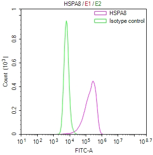
Overlay Peak curve showing THP-1 cells stained with CSB-RA010829MA1HU (red line) at 1:100. The cells were fixed in 4% formaldehyde and permeated by 0.2% TritonX-100 for10min. Then 10% normal goat serum to block non-specific protein-protein interactions followed by the antibody (1ug/1*106cells) for 45min at 4°C. The secondary antibody used was Fluorescein (FITC) AffiniPure Goat Anti-Human IgG, Fcgamma fragment specific at 1:200 dilution for 35min at 4°C.Control antibody (green line) was human IgG1 (1ug/1*106cells) used under the same conditions. Acquisition of >10,000 events was performed. |
|
|
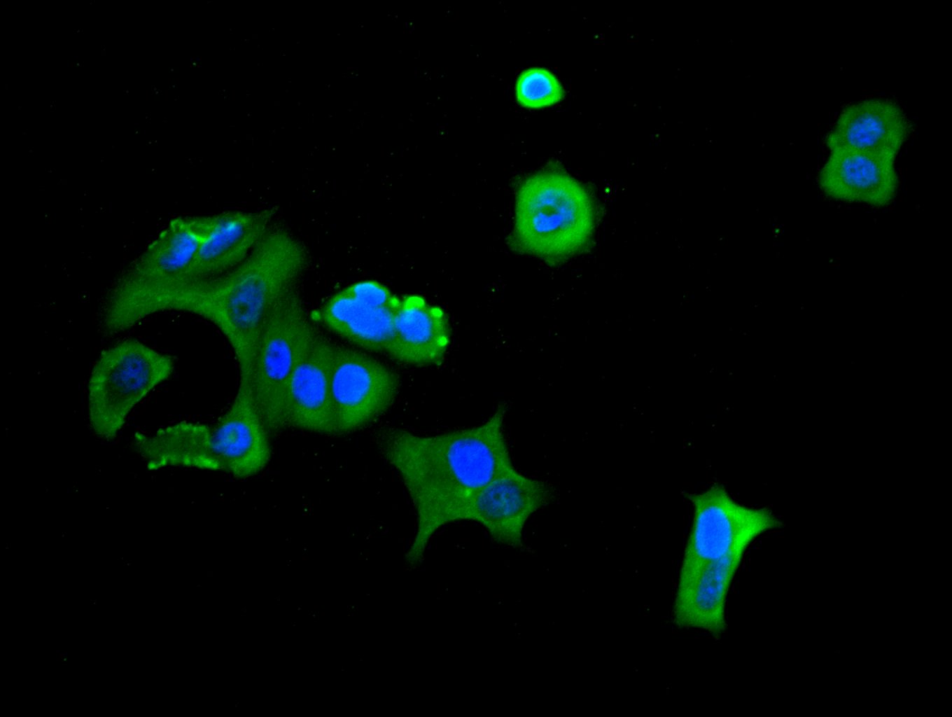
Immunofluorescence staining of MCF-7 cell with CSB-RA010829MA1HU at 1:30 counter-stained with DAPI. The cells were fixed in 4% formaldehyde, permeabilized using 0.2% Triton X-100 and blocked in 10% normal Goat Serum. The cells were then incubated with the antibody overnight at 4C. The secondary antibody was Alexa Fluor 488-congugated AffiniPure Goat Anti-Human IgG(H+L) . |
|
|
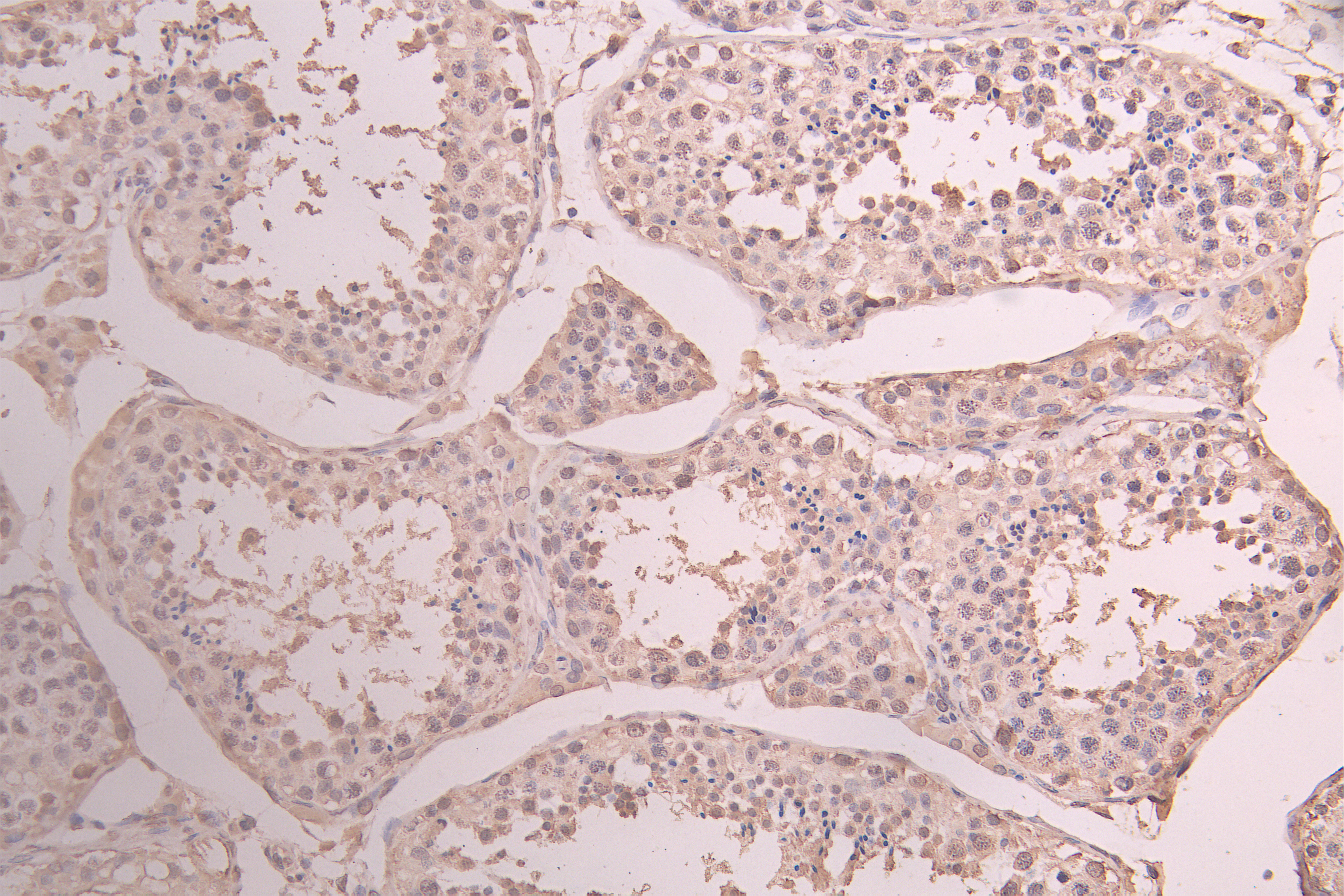
IHC image of CSB-RA010829MA1HU diluted at 1:100 and staining in paraffin-embedded human testis tissue performed on a Leica BondTM system. After dewaxing and hydration, antigen retrieval was mediated by high pressure in a citrate buffer (pH 6.0) . Section was blocked with 10% normal goat serum 30min at RT. Then primary antibody (1% BSA) was incubated at 4C overnight. The primary is detected by a Goat anti-human polymer IgG labeled by HRP and visualized using 0.05% DAB. |
|
|
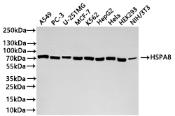
Western Blot Positive WB detected in: A549 whole cell lysate(20µg) , PC-3 whole cell lysate(20µg) , U-251MG whole cell lysate(20µg) , MCF-7 whole cell lysate(20µg) , K562 whole cell lysate(20µg) , HepG2 whole cell lysate(20µg) , HeLa whole cell lysate(20µg) , HEK293 whole cell lysate(20µg) , NIH/3T3 whole cell lysate(20µg) All lanes: HSPA8 antibody at 1:1000 Secondary Goat polyclonal to rabbit IgG at 1/40000 dilution Predicted band size: 71 kDa Observed band size: 71 kDa Exposure time:10s |
|
|
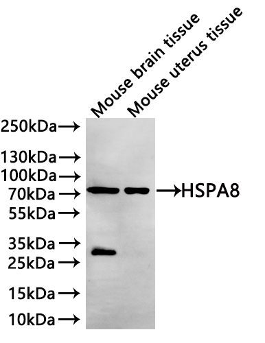
Western Blot Positive WB detected in: Mouse brain tissue lysate(20µg) , Mouse uterus tissue lysate(20µg) All lanes: HSPA8 antibody at 1:1000 Secondary Goat polyclonal to human IgG at 1/40000 dilution Predicted band size: 70.9 kDa Observed band size: 71 kDa Exposure time: 2min |
Product Guarantee and Expert Support
