
HSP90AA1 Recombinant Monoclonal Antibody, Clone: [4B5], Unconjugated, Rabbit
Catalog Number:
CSB-RA011087A0HU
- Images (5)
| Article Name: | HSP90AA1 Recombinant Monoclonal Antibody, Clone: [4B5], Unconjugated, Rabbit |
| Biozol Catalog Number: | CSB-RA011087A0HU |
| Supplier Catalog Number: | CSB-RA011087A0HU |
| Alternative Catalog Number: | CSB-RA011087A0HU-100UL, CSB-RA011087A0HU-50UL |
| Manufacturer: | Cusabio |
| Host: | Rabbit |
| Category: | Antikörper |
| Application: | ELISA, IF, IHC, IP, WB |
| Species Reactivity: | Human, Rat |
| Conjugation: | Unconjugated |
| Alternative Names: | Heat shock protein HSP 90-alpha, Heat shock 86 kDa, HSP 86, HSP86, Lipopolysaccharide-associated protein 2, LAP-2, LPS-associated protein 2, Renal carcinoma antigen NY-REN-38, HSP90AA1, HSP90A, HSPC1, HSPCA |
| Clonality: | Monoclonal |
| Clone Designation: | [4B5] |
| UniProt: | P07900 |
| Buffer: | Rabbit IgG in 10mM phosphate buffered saline , pH 7.4, 150mM sodium chloride, 0.05% BSA, 0.02% sodium azide and 50% glycerol. |
| Purity: | Affinity-chromatography |
| Form: | Liquid |
| Target: | HSP90AA1 |
| Antibody Type: | Recombinant Antibody |
| Application Dilute: | Recommended dilution: WB:1:500-1:5000, IHC:1:50-1:200, IF:1:20-1:200, IP:1:200-1:1000 |
|
|
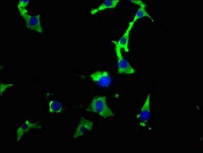
Immunofluorescence staining of NIH/3T3 cells with CSB-RA011087A0HU at 1:26, counter-stained with DAPI. The cells were fixed in 4% formaldehyde, permeabilized using 0.2% Triton X-100 and blocked in 10% normal Goat Serum. The cells were then incubated with the antibody overnight at 4°C. The secondary antibody was Alexa Fluor 488-congugated AffiniPure Goat Anti-Rabbit IgG (H+L) . |
|
|
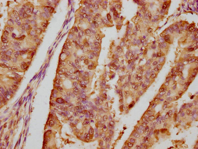
IHC image of CSB-RA011087A0HU diluted at 1:80 and staining in paraffin-embedded human endometrial cancer performed on a Leica BondTM system. After dewaxing and hydration, antigen retrieval was mediated by high pressure in a citrate buffer (pH 6.0) . Section was blocked with 10% normal goat serum 30min at RT. Then primary antibody (1% BSA) was incubated at 4°C overnight. The primary is detected by a biotinylated secondary antibody and visualized using an HRP conjugated SP system. |
|
|
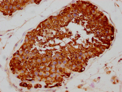
IHC image of CSB-RA011087A0HU diluted at 1:80 and staining in paraffin-embedded human testis tissue performed on a Leica BondTM system. After dewaxing and hydration, antigen retrieval was mediated by high pressure in a citrate buffer (pH 6.0) . Section was blocked with 10% normal goat serum 30min at RT. Then primary antibody (1% BSA) was incubated at 4°C overnight. The primary is detected by a biotinylated secondary antibody and visualized using an HRP conjugated SP system. |
|
|
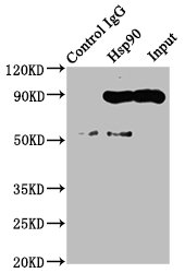
Immunoprecipitating Hsp90 in Hela whole cell lysate Lane 1: Rabbit control IgG instead of CSB-RA011087A0HU in Hela whole cell lysate.For western blotting, a HRP-conjugated Protein G antibody was used as the secondary antibody (1/2000) Lane 2: CSB-RA011087A0HU (3µg) + Hela whole cell lysate (500µg) Lane 3: Hela whole cell lysate (20µg) |
|
|
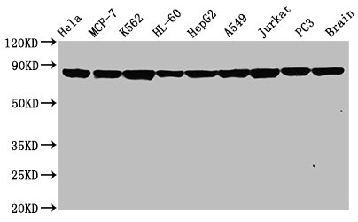
Western Blot Positive WB detected in: Hela whole cell lysate, MCF-7 whole cell lysate, K562 whole cell lysate, HL-60 whole cell lysate, HepG2 whole cell lysate, A549 whole cell lysate, Jurkat whole cell lysate, PC3 whole cell lysate, Rat brain tissue All lanes: Hsp90 alpha antibody at 0.8µg/ml Secondary Goat polyclonal to rabbit IgG at 1/50000 dilution Predicted band size: 85, 99 KDa Observed band size: 85 KDa |
Product Guarantee and Expert Support
