
PRKCD/PRKCQ/Prkcd Recombinant Monoclonal Antibody, Clone: [24F7], Unconjugated, Rabbit
Catalog Number:
CSB-RA057607A0HU
- Images (5)
| Article Name: | PRKCD/PRKCQ/Prkcd Recombinant Monoclonal Antibody, Clone: [24F7], Unconjugated, Rabbit |
| Biozol Catalog Number: | CSB-RA057607A0HU |
| Supplier Catalog Number: | CSB-RA057607A0HU |
| Alternative Catalog Number: | CSB-RA057607A0HU-100UL, CSB-RA057607A0HU-50UL |
| Manufacturer: | Cusabio |
| Host: | Rabbit |
| Category: | Antikörper |
| Application: | ELISA, FC, IF, IHC, WB |
| Species Reactivity: | Human, Mouse, Rat |
| Conjugation: | Unconjugated |
| Alternative Names: | CVID9 antibody, D14Ertd420e antibody, Kinase PKC delta antibody, KPCD antibody, KPCD_HUMAN antibody, MAY 1 antibody, MAY1 antibody, MGC49908 antibody, nPKC delta antibody, nPKC-delta antibody, PCKd antibody, PKC d antibody, PKC delta antibody, PKCD antibody, PKCdelta antibody, PRKC D antibody, PRKC delta antibody, Prkcd antibody, Protein Kinase C delta antibody, Protein kinase C delta type antibody, Protein kinase C delta VIII antibody, Protein Kinase Cdelta antibody, Tyrosine protein kinase PRKCD antibody, KPCT_HUMAN antibody, MGC126514 antibody, MGC141919 antibody, nPKC theta antibody, nPKC-theta antibody, nPKCtheta antibody, OTTHUMP00000043364 antibody, OTTHUMP00000043365 antibody, PKC 0 antibody, PKC0 antibody, Prkcq antibody, PRKCT antibody, Protein kinase C theta antibody, Protein kinase C theta type antibody, Protein Kinase Ctheta antibody,Prkcd, PkcdProtein kinase C delta type, EC 2.7.11.13, Tyrosine-protein kinase PRKCD, EC 2.7.10.2, nPKC-delta) [Cleaved into: Protein kinase C delta type regulatory subunit, Protein kinase C delta type catalytic subunit, Sphingosine-dependent protein kinase-1, SDK1)] |
| Clonality: | Monoclonal |
| Clone Designation: | [24F7] |
| UniProt: | Q05655 |
| Buffer: | Rabbit IgG in 10mM phosphate buffered saline , pH 7.4, 150mM sodium chloride, 0.05% BSA, 0.02% sodium azide and 50% glycerol. |
| Purity: | Affinity-chromatography |
| Form: | Liquid |
| Target: | PRKCD/PRKCQ/Prkcd |
| Antibody Type: | Recombinant Antibody |
| Application Dilute: | Recommended dilution: WB:1:500-1:5000, IHC:1:50-1:200, IF:1:50-1:200, FC:1:50-1:200 |
|
|
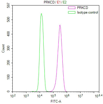
Overlay Peak curve showing A431 cells stained with CSB-RA057607A0HU (red line) at 1:100. The cells were fixed in 4% formaldehyde and permeated by 0.2% TritonX-100 for10min. Then 10% normal goat serum to block non-specific protein-protein interactions followed by the antibody (1ug/1*106cells) for 45min at 4°C. The secondary antibody used was FITC-conjugated goat anti-rabbit IgG (H+L) at 1/200 dilution for 35min at 4°C.Control antibody (green line) was Rabbit IgG (1ug/1*106cells) used under the same conditions. Acquisition of >10, 000 events was performed. |
|
|
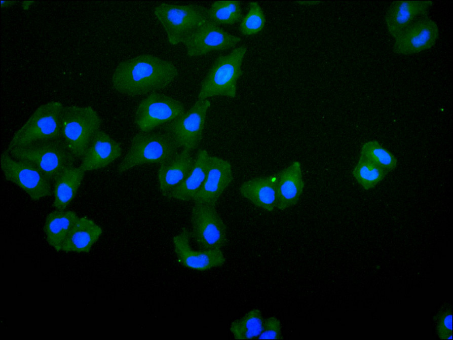
Immunofluorescence staining of HeLa cell with CSB-RA057607A0HU at 1:50 , counter-stained with DAPI. The cells were fixed in 4% formaldehyde, permeabilized using 0.2% Triton X-100 and blocked in 10% normal Goat Serum. The cells were then incubated with the antibody overnight at 4C. The secondary antibody was Alexa Fluor 488-congugated AffiniPure Goat Anti-Rabbit IgG(H+L). |
|
|
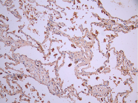
IHC image of CSB-RA057607A0HU diluted at 1:100 and staining in paraffin-embedded human lung tissue performed on a Leica BondTM system. After dewaxing and hydration, antigen retrieval was mediated by high pressure in a citrate buffer (pH 6.0). Section was blocked with 10% normal goat serum 30min at RT. Then primary antibody (1% BSA) was incubated at 4C overnight. The primary is detected by a Goat anti-rabbit polymer IgG labeled by HRP and visualized using 0.05% DAB. |
|
|
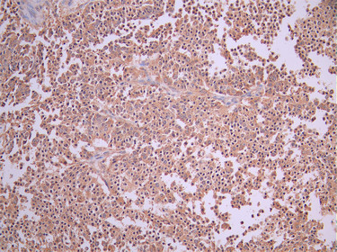
IHC image of CSB-RA057607A0HU diluted at 1:100 and staining in paraffin-embedded human liver cancer performed on a Leica BondTM system. After dewaxing and hydration, antigen retrieval was mediated by high pressure in a citrate buffer (pH 6.0). Section was blocked with 10% normal goat serum 30min at RT. Then primary antibody (1% BSA) was incubated at 4C overnight. The primary is detected by a Goat anti-rabbit polymer IgG labeled by HRP and visualized using 0.05% DAB. |
|
|
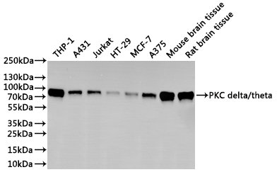
Western Blot Positive WB detected in: THP-1 whole cell lysate(30µg), A431 whole cell lysate(30µg), Jurkat whole cell lysate(30µg), HT-29 whole cell lysate(30µg), MCF7 whole cell lysate(30µg), A375 whole cell lysate(30µg), Mouse brain tissue lysate(30µg), Rat brain tissue lysate(30µg) All lanes: PKC delta/theta antibody at 1:1000 Secondary Goat polyclonal to rabbit IgG at 1/40000 dilution Predicted band size: 78 kDa Observed band size: 78 kDa Exposure time:2min |
Product Guarantee and Expert Support
