The protein encoded by this gene is a DNA-binding transcription factor that activates many muscle-specific, growth factor-induced, and stress-induced genes. The encoded protein can act as a homodimer or as a heterodimer and is involved in several cellular processes, including muscle development, neuronal differentiation, cell growth control, and apoptosis. Defects in this gene could be a cause of autosomal dominant coronary artery disease 1 with myocardial infarction (ADCAD1). Several transcript variants encoding different isoforms have been found for this gene.[provided by RefSeq, Jan 2010],
Western Blot: 1/500 - 1/2000. Immunohistochemistry: 1/100 - 1/300. ELISA: 1/20000. Not yet tested in other applications.
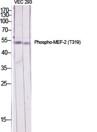
Western Blot analysis of various cells using Phospho-MEF-2 (T319) Polyclonal Antibody diluted at 1:2000 cells nucleus extracted by Minute TM Cytoplasmic and Nuclear Fractionation kit (SC-003,Inventbiotech,MN,USA).
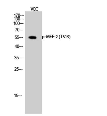
Western Blot analysis of VEC cells using Phospho-MEF-2 (T319) Polyclonal Antibody diluted at 1:2000 cells nucleus extracted by Minute TM Cytoplasmic and Nuclear Fractionation kit (SC-003,Inventbiotech,MN,USA).
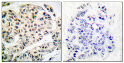
Immunohistochemistry analysis of paraffin-embedded human breast carcinoma, using MEF2A (Phospho-Thr319) Antibody. The picture on the right is blocked with the phospho peptide.
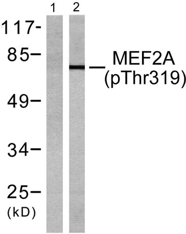
Western blot analysis of lysates from K562 cells treated with UV 15, using MEF2A (Phospho-Thr319) Antibody. The lane on the left is blocked with the phospho peptide.
* VAT and and shipping costs not included. Errors and price changes excepted