The antiserum was produced against synthesized peptide derived from human RFA2 around the phosphorylation site of Ser33. AA range:1-50
Alternative Names:
RPA2, REPA2, RPA32, RPA34, Replication protein A 32 kDa subunit, RP-A p32, Replication factor A protein 2, RF-A protein 2, Replication protein A 34 kDa subunit, RP-A p34
function:Required for DNA recombination, repair and replication. The activity of RP-A is mediated by single-stranded DNA binding and protein interactions.,PTM:Phosphorylated in a cell-cycle-dependent manner (from the S phase until mitosis). Phosphorylated by ATR upon DNA damage, which promotes its translocation to nuclear foci. Can be phosphorylated in vitro by PRKDC/DNA-PK in the presence of Ku and DNA, and by CDC2.,subcellular location:Also present in PML nuclear bodies. Redistributes to discrete nuclear foci upon DNA damage.,subunit:Heterotrimer of 70, 32 and 14 kDa chains. The DNA-binding activity may reside exclusively on the 70 kDa subunit. Binds to SERTAD3/RBT1. Interacts with TIPIN.,
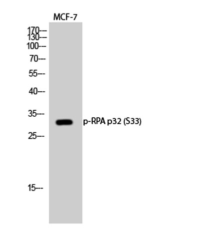
Western Blot analysis of various cells using Phospho-RPA p32 (S33) Polyclonal Antibody diluted at 1:500 cells nucleus extracted by Minute TM Cytoplasmic and Nuclear Fractionation kit (SC-003,Inventbiotech,MN,USA).
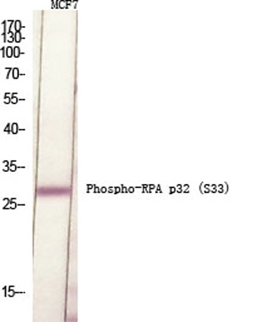
Western Blot analysis of MCF-7 cells using Phospho-RPA p32 (S33) Polyclonal Antibody diluted at 1:500 cells nucleus extracted by Minute TM Cytoplasmic and Nuclear Fractionation kit (SC-003,Inventbiotech,MN,USA).
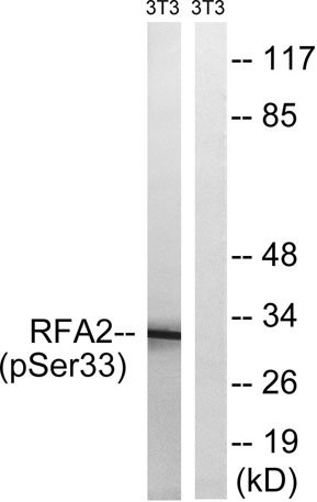
Western blot analysis of lysates from NIH/3T3 cells treated with Adriamycin 0.5ug/ml 24h, using RFA2 (Phospho-Ser33) Antibody. The lane on the right is blocked with the phospho peptide.
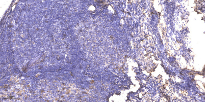
Immunohistochemical analysis of paraffin-embedded human cervical carcinoma. 1, Antibody was diluted at 1:200(4 overnight). 2, Tris-EDTA,pH9.0 was used for antigen retrieval. 3,Secondary antibody was diluted at 1:200(room temperature, 45min).
* VAT and and shipping costs not included. Errors and price changes excepted