The antiserum was produced against synthesized peptide derived from human KSR around the phosphorylation site of Ser392. AA range:358-407
Alternative Names:
KSR1, KSR, Kinase suppressor of Ras 1
caution:The sequence shown here is derived from an Ensembl automatic analysis pipeline and should be considered as preliminary data.,function:Location-regulated scaffolding protein connecting MEK to RAF. Promotes MEK and RAF phosphorylation and activity through assembly of an activated signaling complex. By itself, it has no demonstrated kinase activity.,PTM:Phosphorylated on Ser-309 and, to a higher extent, on Ser-404 by MARK3. Dephosphorylated on Ser-404 by PPP2CA. In resting cells, phosphorylated KSR1 is cytoplasmic and in stimulated cells, dephosphorylated KSR1 is membrane-associated.,similarity:Belongs to the protein kinase superfamily. TKL Ser/Thr protein kinase family.,similarity:Contains 1 phorbol-ester/DAG-type zinc finger.,similarity:Contains 1 protein kinase domain.,subcellular location:In unstimulated cells, where the phosphorylated form is bound to a 14-3-3 protein, sequestration in the cytoplasm occurs. Following growth factor treatment, the protein is free for membrane translocation, and it moves from the cytoplasm to the cell periphery.,subunit:Interacts with HSPCA/HSP90, YWHAB/14-3-3, CDC37, MAP2K/MEK, MARK3, PPP2R1A and PPP2CA. Also interacts with RAF and MAPK/ERK, in a Ras-dependent manner (By similarity). The binding of 14-3-3 proteins to phosphorylated KSR prevents the membrane localization.,
Western Blot: 1/500 - 1/2000. Immunohistochemistry: 1/100 - 1/300. ELISA: 1/20000. Not yet tested in other applications.
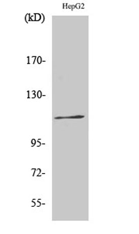
Western Blot analysis of various cells using Phospho-Ksr-1 (S392) Polyclonal Antibody
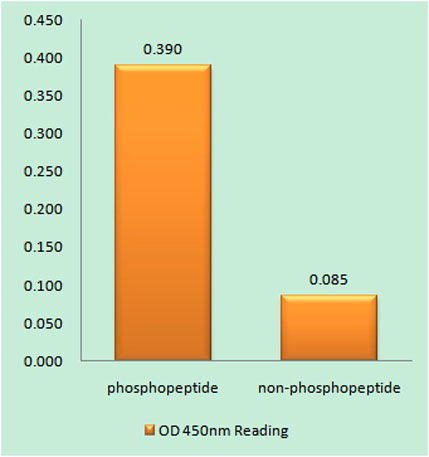
Enzyme-Linked Immunosorbent Assay (Phospho-ELISA) for Immunogen Phosphopeptide (Phospho-left) and Non-Phosphopeptide (Phospho-right), using KSR (Phospho-Ser392) Antibody
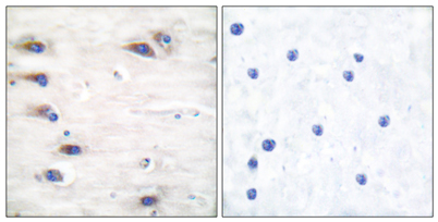
Immunohistochemistry analysis of paraffin-embedded human brain, using KSR (Phospho-Ser392) Antibody. The picture on the right is blocked with the phospho peptide.
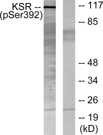
Western blot analysis of lysates from HepG2 cells, using KSR (Phospho-Ser392) Antibody. The lane on the right is blocked with the phospho peptide.
* VAT and and shipping costs not included. Errors and price changes excepted