The antiserum was produced against synthesized peptide derived from human AKAP5. AA range:1-50
Alternative Names:
AKAP5, AKAP79, A-kinase anchor protein 5, AKAP-5, A-kinase anchor protein 79 kDa, AKAP 79, H21, cAMP-dependent protein kinase regulatory subunit II high affinity-binding protein
The A-kinase anchor proteins (AKAPs) are a group of structurally diverse proteins, which have the common function of binding to the regulatory subunit of protein kinase A (PKA) and confining the holoenzyme to discrete locations within the cell. This gene encodes a member of the AKAP family. The encoded protein binds to the RII-beta regulatory subunit of PKA, and also to protein kinase C and the phosphatase calcineurin. It is predominantly expressed in cerebral cortex and may anchor the PKA protein at postsynaptic densities (PSD) and be involved in the regulation of postsynaptic events. It is also expressed in T lymphocytes and may function to inhibit interleukin-2 transcription by disrupting calcineurin-dependent dephosphorylation of NFAT. [provided by RefSeq, Jul 2008],
Western Blot: 1/500 - 1/2000. Immunohistochemistry: 1/100 - 1/300. Immunofluorescence: 1/200 - 1/1000. ELISA: 1/10000. Not yet tested in other applications.
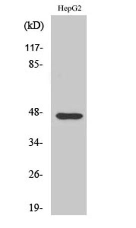
Western Blot analysis of various cells using AKAP 79 Polyclonal Antibody diluted at 1:1000
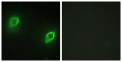
Immunofluorescence analysis of HeLa cells, using AKAP5 Antibody. The picture on the right is blocked with the synthesized peptide.
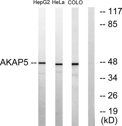
Western blot analysis of lysates from HepG2, HeLa, and COLO205 cells, using AKAP5 Antibody. The lane on the right is blocked with the synthesized peptide.
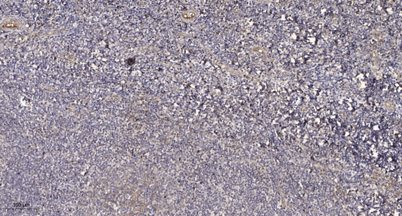
Immunohistochemical analysis of paraffin-embedded human tonsil. 1, Antibody was diluted at 1:200(4 overnight). 2, Tris-EDTA,pH9.0 was used for antigen retrieval. 3,Secondary antibody was diluted at 1:200(room temperature, 30min).
* VAT and and shipping costs not included. Errors and price changes excepted