
LOXL2 antibody, Unconjugated, Rabbit, Polyclonal
Catalog Number:
GTX105085
- Images (8)

| Article Name: | LOXL2 antibody, Unconjugated, Rabbit, Polyclonal |
| Biozol Catalog Number: | GTX105085 |
| Supplier Catalog Number: | GTX105085 |
| Alternative Catalog Number: | GTX105085-100,GTX105085-25 |
| Manufacturer: | GeneTex |
| Host: | Rabbit |
| Category: | Antikörper |
| Application: | ELISA, ICC, IHC-P, WB |
| Species Reactivity: | Human, Mouse |
| Immunogen: | Recombinant protein encompassing a sequence within the center region of human LOXL2. The exact sequence is proprietary. |
| Conjugation: | Unconjugated |
| Alternative Names: | lysyl oxidase like 2 , LOR , LOR2 , WS9-14 |
| Clonality: | Polyclonal |
| Concentration: | 0.86 mg/ml (Please refer to the vial label for the specific concentration.) |
| Molecular Weight: | 87 |
| Sensitivity: | Mouse reactivity has been validated by IHC-P only, not yet been tested by other applications. |
| NCBI: | 4017 |
| UniProt: | Q9Y4K0 |
| Buffer: | 1XPBS, 20% Glycerol (pH7), 0.025% ProClin 300. |
| Purity: | Purified by antigen-affinity chromatography. |
| Form: | Liquid |
| Application Notes: | WB: 1:500-1:10000. ICC/IF: 1:100-1:1000. IHC-P: 1:100-1:1000. *Optimal dilutions/concentrations should be determined by the researcher.Not tested in other applications. |
|
|
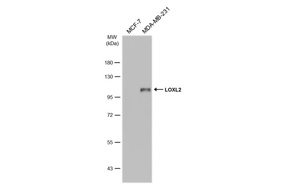
GTX105085 WB Image |
|
|
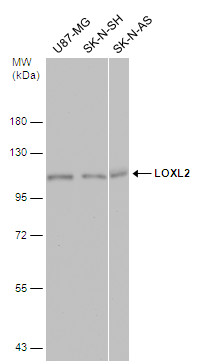
Various whole cell extracts (30 μg) were separated by 7.5% SDS-PAGE, and the membrane was blotted with LOXL2 antibody (GTX105085) diluted at 1:5000. The HRP-conjugated anti-rabbit IgG antibody (GTX213110-01) was used to detect the primary antibody. |
|
|
LOXL2 antibody detects LOXL2 protein at nucleus by immunohistochemical analysis.Sample: Paraffin-embedded human esophageal carcinoma.LOXL2 stained by LOXL2 antibody (GTX105085) diluted at 1:3000.Antigen Retrieval: Citrate buffer, pH 6.0, 15 min |
|
|
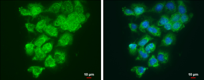
LOXL2 antibody detects LOXL2 protein at cytoplasm and nucleus by immunofluorescent analysis. Sample: A431 cells were fixed in ice-cold MeOH for 5 min. Green: LOXL2 protein stained by LOXL2 antibody (GTX105085) diluted at 1:500.. Blue: Hoechst 33342 staining. Scale bar = 10 μm. |
|
|
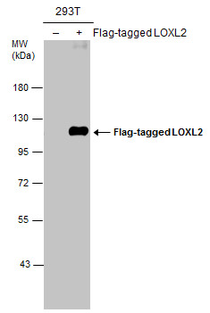
Non-transfected (–) and transfected (+) 293T whole cell extracts (30 μg) were separated by 7.5% SDS-PAGE, and the membrane was blotted with LOXL2 antibody (GTX105085) diluted at 1:5000. |
|
|
Various whole cell extracts (30 μg) were separated by 7.5% SDS-PAGE, and the membrane was blotted with LOXL2 antibody (GTX105085) diluted at 1:1000. The HRP-conjugated anti-rabbit IgG antibody (GTX213110-01) was used to detect the primary antibody. |
|
|
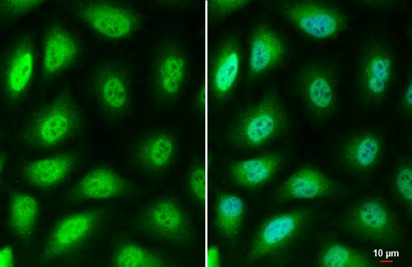
LOXL2 antibody detects LOXL2 protein by immunofluorescent analysis.Sample: HeLa cells were fixed in 4% paraformaldehyde at RT for 15 min.Green: LOXL2 stained by LOXL2 antibody (GTX105085) diluted at 1:500.Blue: Hoechst 33342 staining.Scale bar= 10 μm. |
|
|
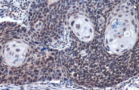
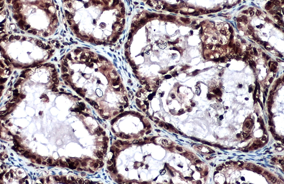
LOXL2 antibody detects LOXL2 protein at cytoplasm and nucleus by immunohistochemical analysis.Sample: Paraffin-embedded human ovarian cancer.LOXL2 stained by LOXL2 antibody (GTX105085) diluted at 1:500.Antigen Retrieval: Citrate buffer, pH 6.0, 15 min |
Product Guarantee and Expert Support
