
Mre11 antibody [C1C3-2], Unconjugated, Rabbit, Polyclonal
Catalog Number:
GTX111814
- Images (5)
| Article Name: | Mre11 antibody [C1C3-2], Unconjugated, Rabbit, Polyclonal |
| Biozol Catalog Number: | GTX111814 |
| Supplier Catalog Number: | GTX111814 |
| Alternative Catalog Number: | GTX111814-100,GTX111814-25 |
| Manufacturer: | GeneTex |
| Host: | Rabbit |
| Category: | Antikörper |
| Application: | ICC, IP, WB |
| Species Reactivity: | Human |
| Immunogen: | Recombinant protein encompassing a sequence within the C-terminus region of human Mre11. The exact sequence is proprietary. |
| Conjugation: | Unconjugated |
| Alternative Names: | MRE11 homolog, double strand break repair nuclease , ATLD , HNGS1 , MRE11A , MRE11B |
| Application Notes: | WB: 1:500-1:3000. ICC/IF: 1:100-1:1000. IP: 1:100-1:500. *Optimal dilutions/concentrations should be determined by the researcher.Not tested in other applications. |
|
|
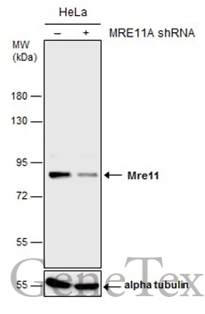
GTX111814 WB Image |
|
|
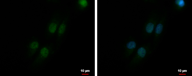
Mre11 antibody [C1C3-2] detects Mre11 protein at nucleus by immunofluorescent analysis. Sample: SKNSH cells were fixed in 4% paraformaldehyde at RT for 15 min. Green: Mre11 protein stained by Mre11 antibody [C1C3-2] (GTX111814) diluted at 1:500. Blue: Hoechst 33342 staining. Scale bar = 10 μm. |
|
|
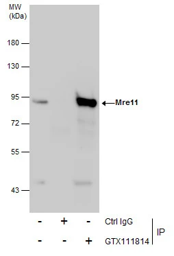
Immunoprecipitation of Mre11 protein from HeLa whole cell extracts using 5 μg of Mre11 antibody [C1C3-2] (GTX111814). Western blot analysis was performed using Mre11 antibody [C1C3-2] (GTX111814). EasyBlot anti-Rabbit IgG (GTX221666-01) was used as a secondary reagent. |
|
|
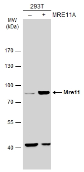
Non-transfected (–) and transfected (+) 293T whole cell extracts (30 μg) were separated by 7.5% SDS-PAGE, and the membrane was blotted with Mre11 antibody [C1C3-2] (GTX111814) diluted at 1:1000. The HRP-conjugated anti-rabbit IgG antibody (GTX213110-01) was used to detect the primary antibody. |
|
|
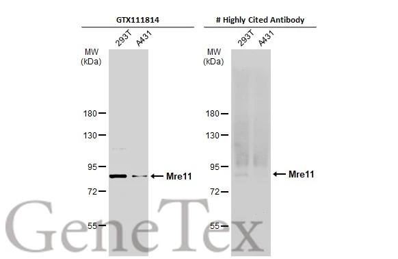
Various whole cell extracts (30 μg) were separated by 7.5% SDS-PAGE, and the membranes were blotted with Mre11 antibody [C1C3-2] (GTX111814) diluted at 1:1000 and competitor's antibody diluted at 1:1000. The HRP-conjugated anti-rabbit IgG antibody (GTX213110-01) was used to detect the primary antibody. *The competitor is not affiliated with GeneTex and does not endorse this product. |
Product Guarantee and Expert Support
