
PRX III antibody [N1C3], Unconjugated, Rabbit, Polyclonal
Catalog Number:
GTX112004
- Images (5)
| Article Name: | PRX III antibody [N1C3], Unconjugated, Rabbit, Polyclonal |
| Biozol Catalog Number: | GTX112004 |
| Supplier Catalog Number: | GTX112004 |
| Alternative Catalog Number: | GTX112004-100,GTX112004-25 |
| Manufacturer: | GeneTex |
| Host: | Rabbit |
| Category: | Antikörper |
| Application: | ICC, IHC-P, WB |
| Species Reactivity: | Human, Mouse |
| Immunogen: | Recombinant protein encompassing a sequence within the center region of human PRX III. The exact sequence is proprietary. |
| Conjugation: | Unconjugated |
| Alternative Names: | peroxiredoxin 3 , AOP-1 , AOP1 , HBC189 , MER5 , PRO1748 , SP-22 , prx-III |
| Application Notes: | WB: 1:500-1:3000. ICC/IF: 1:100-1:1000. IHC-P: 1:100-1:1000. *Optimal dilutions/concentrations should be determined by the researcher.Not tested in other applications. |
|
|
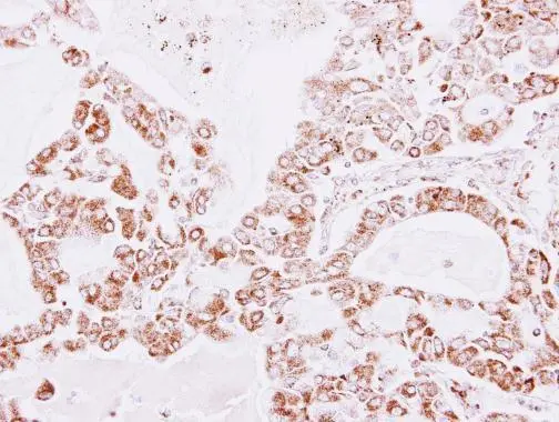
GTX112004 IHC-P Image |
|
|
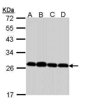
Sample (30 ug of whole cell lysate) A: Hela B: Hep G2 (GTX27900) C: Molt-4 (GTX27912) D: Raji 12% SDS PAGE GTX112004 diluted at 1:1000 |
|
|
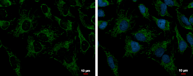
Peroxiredoxin 3 antibody [N1C3] detects Peroxiredoxin 3 protein at mitochondria by immunofluorescent analysis. Sample: HeLa cells were fixed in 2% paraformaldehyde/culture medium at 37oC for 30 min. Green: Peroxiredoxin 3 protein stained by Peroxiredoxin 3 antibody [N1C3] (GTX112004) diluted at 1:1000. Blue: Hoechst 33342 staining. |
|
|
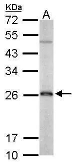
Sample (50 ug of whole cell lysate) A: mouse brain 12% SDS PAGE GTX112004 diluted at 1:1000 |
|
|
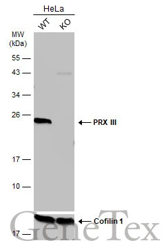
Wild-type (WT) and PRX III knockout (KO) HeLa cell extracts (30 μg) were separated by 12% SDS-PAGE, and the membrane was blotted with PRX III antibody [N1C3] (GTX112004) diluted at 1:1000. The HRP-conjugated anti-rabbit IgG antibody (GTX213110-01) was used to detect the primary antibody. |
Product Guarantee and Expert Support
