
GLUD1 + GLUD2 antibody, Unconjugated, Rabbit, Polyclonal
Catalog Number:
GTX112361
- Images (5)
| Article Name: | GLUD1 + GLUD2 antibody, Unconjugated, Rabbit, Polyclonal |
| Biozol Catalog Number: | GTX112361 |
| Supplier Catalog Number: | GTX112361 |
| Alternative Catalog Number: | GTX112361-100,GTX112361-25 |
| Manufacturer: | GeneTex |
| Host: | Rabbit |
| Category: | Antikörper |
| Application: | ICC, IHC-P, WB |
| Species Reactivity: | Drosophila, Human, Mouse, Rat |
| Immunogen: | Recombinant protein encompassing a sequence within the center region of human GLUD1+2. The exact sequence is proprietary. |
| Conjugation: | Unconjugated |
| Alternative Names: | GDH2 antibody , GLUDP1 antibody , GLUD2 antibody , GDH 2 antibody , glutamate dehydrogenase 2 , mitochondrial antibody , glutamate dehydrogenase pseudogene 1 antibody , glutamate dehydrogenase 2 antibody |
| Clonality: | Polyclonal |
| Concentration: | 1 mg/ml (Please refer to the vial label for the specific concentration.) |
| Molecular Weight: | 61 |
| NCBI: | 2747 |
| Buffer: | 1XPBS (pH7), 1% BSA, 20% Glycerol, 0.01% Thimerosal. |
| Purity: | Purified by antigen-affinity chromatography. |
| Form: | Liquid |
| Application Notes: | WB: 1:500-1:3000. ICC/IF: 1:100-1:1000. IHC-P: 1:100-1:1000. *Optimal dilutions/concentrations should be determined by the researcher.Not tested in other applications. |
|
|
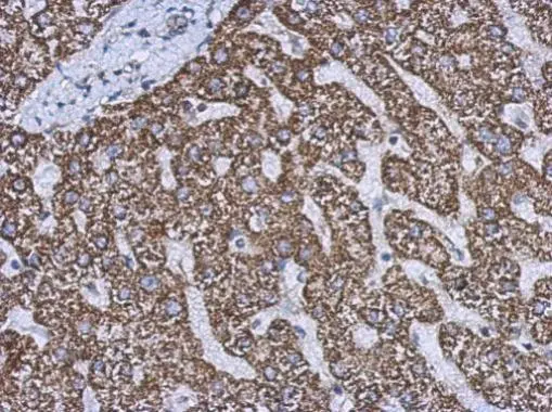
GTX112361 IHC-P Image |
|
|
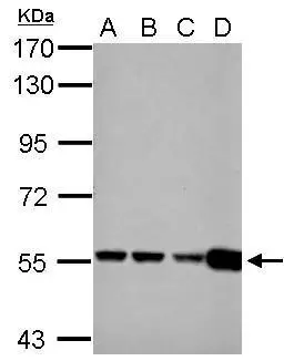
Sample (30 μg of whole cell lysate) A: 293T B: A431 C: HeLa D: HepG2 7.5% SDS PAGE GTX112361 diluted at 1:1000 The HRP-conjugated anti-rabbit IgG antibody (GTX213110-01) was used to detect the primary antibody. |
|
|
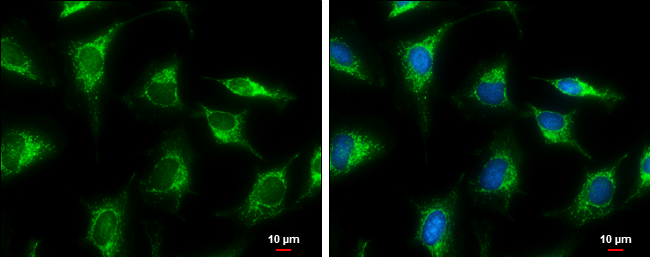
GLUD1+2 antibody detects GLUD1+2 protein at mitochondria by immunofluorescent analysis. Sample: HeLa cells were fixed in ice-cold MeOH for 5 min. Green: GLUD1+2 protein stained by GLUD1+2 antibody (GTX112361) diluted at 1:500. Blue: Hoechst 33342 staining. Scale bar = 10 μm. |
|
|
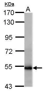
Sample (50 μg of whole cell lysate) A: mouse testis 7.5% SDS PAGE GTX112361 diluted at 1:1000 The HRP-conjugated anti-rabbit IgG antibody (GTX213110-01) was used to detect the primary antibody. |
|
|
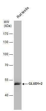
Rat tissue extract (50 μg) was separated by 7.5% SDS-PAGE, and the membrane was blotted with GLUD1+2 antibody (GTX112361) diluted at 1:1000. The HRP-conjugated anti-rabbit IgG antibody (GTX213110-01) was used to detect the primary antibody. |
Product Guarantee and Expert Support
