
HDAC3 antibody, Unconjugated, Rabbit, Polyclonal
Catalog Number:
GTX113303
- Images (9)
| Article Name: | HDAC3 antibody, Unconjugated, Rabbit, Polyclonal |
| Biozol Catalog Number: | GTX113303 |
| Supplier Catalog Number: | GTX113303 |
| Alternative Catalog Number: | GTX113303-100,GTX113303-25 |
| Manufacturer: | GeneTex |
| Host: | Rabbit |
| Category: | Antikörper |
| Application: | ChIP, ICC, IHC, IHC-P, IP, WB |
| Species Reactivity: | Human, Mouse, Rat, Zebrafish |
| Immunogen: | Recombinant protein encompassing a sequence within the center region of human HDAC3. The exact sequence is proprietary. |
| Conjugation: | Unconjugated |
| Alternative Names: | histone deacetylase 3 , HD3 , KDAC3 , RPD3 , RPD3-2 |
| Application Notes: | WB: 1:500-1:3000. ICC/IF: 1:100-1:1000. IHC-P: 1:100-1:1000. IP: 1:100-1:500. *Optimal dilutions/concentrations should be determined by the researcher.Not tested in other applications. |
|
|
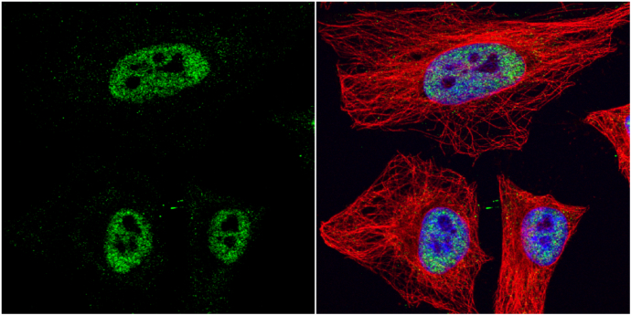
GTX113303 ICC/IF Image |
|
|
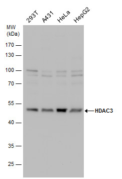
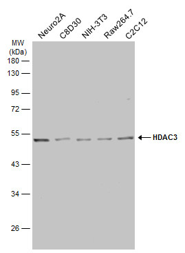
HDAC3 antibody detects HDAC3 protein by Western blot analysis. Various whole cell extracts (30 μg) were separated by 10% SDS-PAGE, and the membrane was blotted with HDAC3 antibody (GTX113303) diluted by 1:1000. |
|
|
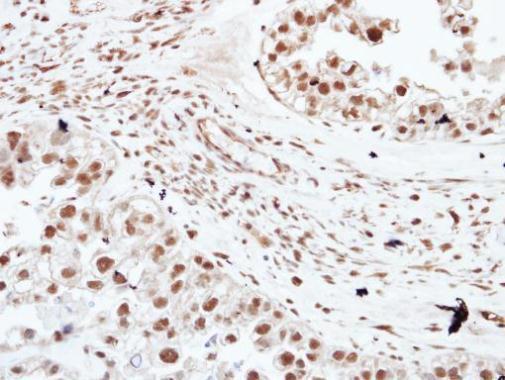
Immunohistochemical analysis of paraffin-embedded human ovarian cancer, using HDAC3(GTX113303) antibody at 1:250 dilution. Antigen Retrieval: Citrate buffer, pH 6.0, 15 min |
|
|
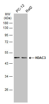
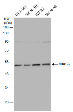
Various whole cell extracts (30 μg) were separated by 10% SDS-PAGE, and the membrane was blotted with HDAC3 antibody (GTX113303) diluted at 1:1000. The HRP-conjugated anti-rabbit IgG antibody (GTX213110-01) was used to detect the primary antibody. |
|
|
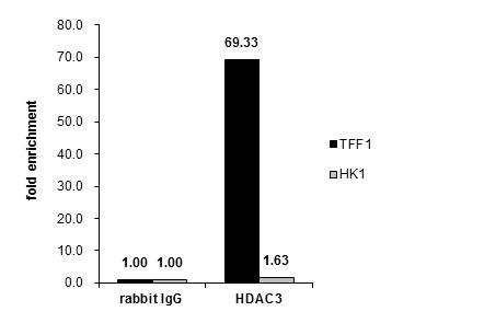
Cross-linked ChIP was performed with MCF-7 chromatin extract and 5 μg of either control rabbit IgG or anti-HDAC3 antibody. The precipitated DNA was detected by PCR with primer set targeting to TFF1 or HK1. |
|
|
Various whole cell extracts (30 μg) were separated by 10% SDS-PAGE, and the membrane was blotted with HDAC3 antibody (GTX113303) diluted at 1:1000. The HRP-conjugated anti-rabbit IgG antibody (GTX213110-01) was used to detect the primary antibody. |
|
|
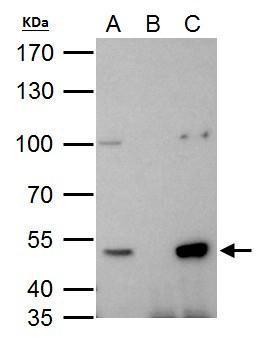
HDAC3 antibody immunoprecipitates HDAC3 protein in IP experiments. IP Sample: 293T whole cell lysate/extract A. 40 μg 293T whole cell lysate/extract B. Control with 2 μg of preimmune rabbit IgG C. Immunoprecipitation of HDAC3 protein by 2 μg of HDAC3 antibody (GTX113303) 7.5% SDS-PAGE The immunoprecipitated HDAC3 protein was detected by HDAC3 antibody (GTX113303) diluted at 1:1000. EasyBlot anti-rabbit IgG (GTX221666-01) was used as a secondary reagent. |
|
|
Various whole cell extracts (30 μg) were separated by 10% SDS-PAGE, and the membrane was blotted with HDAC3 antibody (GTX113303) diluted at 1:500. The HRP-conjugated anti-rabbit IgG antibody (GTX213110-01) was used to detect the primary antibody. |
|
|
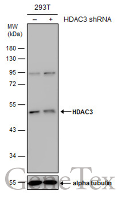
Non-transfected (–) and transfected (+) 293T whole cell extracts (30 μg) were separated by 10% SDS-PAGE, and the membrane was blotted with HDAC3 antibody (GTX113303) diluted at 1:500. The HRP-conjugated anti-rabbit IgG antibody (GTX213110-01) was used to detect the primary antibody. |
Product Guarantee and Expert Support
