
SDHB antibody, Unconjugated, Rabbit, Polyclonal
Catalog Number:
GTX113833
- Images (9)
| Article Name: | SDHB antibody, Unconjugated, Rabbit, Polyclonal |
| Biozol Catalog Number: | GTX113833 |
| Supplier Catalog Number: | GTX113833 |
| Alternative Catalog Number: | GTX113833-100,GTX113833-25 |
| Manufacturer: | GeneTex |
| Host: | Rabbit |
| Category: | Antikörper |
| Application: | ICC, IHC-P, WB |
| Species Reactivity: | Human, Mouse, Rat |
| Immunogen: | Recombinant protein encompassing a sequence within the center region of human SDHB. The exact sequence is proprietary. |
| Conjugation: | Unconjugated |
| Alternative Names: | succinate dehydrogenase complex iron sulfur subunit B , CWS2 , IP , PGL4 , SDH , SDH1 , SDH2 , SDHIP |
| Application Notes: | WB: 1:500-1:3000. ICC/IF: 1:100-1:1000. IHC-P: 1:100-1:1000. *Optimal dilutions/concentrations should be determined by the researcher.Not tested in other applications. |
|
|
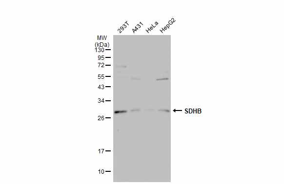
GTX113833 WB Image |
|
|
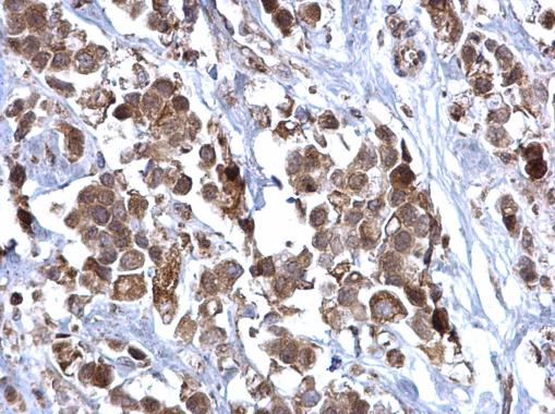
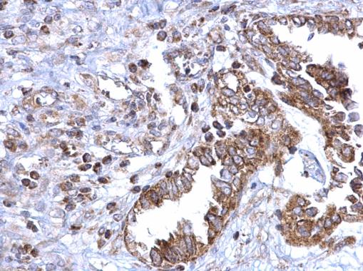
SDHB antibody detects SDHB protein at mitochondria on human colon carcinoma by immunohistochemical analysis. Sample: Paraffin-embedded human colon carcinoma. SDHB antibody (GTX113833) dilution: 1:500. Antigen Retrieval: Trilogy™ (EDTA based, pH 8.0) buffer, 15min |
|
|
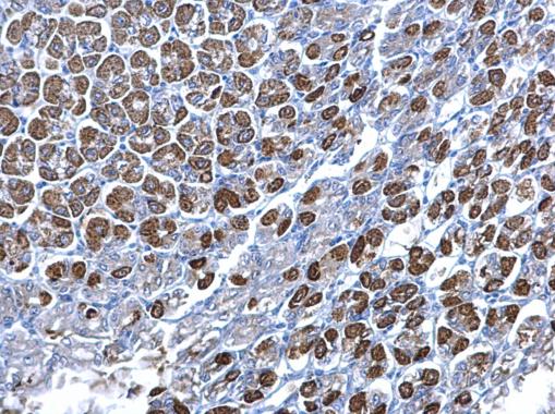
SDHB antibody detects SDHB protein at mitochondria on mouse stomach by immunohistochemical analysis. Sample: Paraffin-embedded mouse stomach. SDHB antibody (GTX113833) dilution: 1:500. Antigen Retrieval: Trilogy™ (EDTA based, pH 8.0) buffer, 15min |
|
|
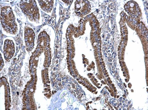
SDHB antibody detects SDHB protein at mitochondria on mouse intestine by immunohistochemical analysis. Sample: Paraffin-embedded mouse intestine. SDHB antibody (GTX113833) dilution: 1:500. _x000D_ Antigen Retrieval: Trilogy™ (EDTA based, pH 8.0) buffer, 15min |
|
|
SDHB antibody detects SDHB protein at mitochondria on human breast carcinoma by immunohistochemical analysis. Sample: Paraffin-embedded human breast carcinoma. SDHB antibody (GTX113833) dilution: 1:500. Antigen Retrieval: Trilogy™ (EDTA based, pH 8.0) buffer, 15min |
|
|
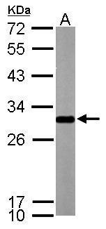
Sample (50 μg of whole cell lysate) A: mouse brain 12% SDS PAGE GTX113833 diluted at 1:1000 The HRP-conjugated anti-rabbit IgG antibody (GTX213110-01) was used to detect the primary antibody. |
|
|
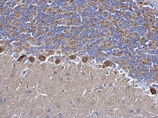
SDHB antibody detects SDHB protein at mitochondria on rat hind brain by immunohistochemical analysis. Sample: Paraffin-embedded rat hind brain. SDHB antibody (GTX113833) dilution: 1:500. Antigen Retrieval: Trilogy™ (EDTA based, pH 8.0) buffer, 15min |
|
|
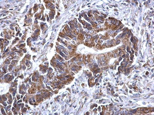
SDHB antibody detects SDHB protein at mitochondria on human ovarian carcinoma by immunohistochemical analysis. Sample: Paraffin-embedded human ovarian carcinoma. SDHB antibody (GTX113833) dilution: 1:500. Antigen Retrieval: Trilogy™ (EDTA based, pH 8.0) buffer, 15min |
|
|
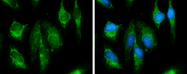
SDHB antibody detects SDHB protein at mitochondria by immunofluorescent analysis. Sample: HeLa cells were fixed in MeOH for 5 min. Green: SDHB protein stained by SDHB antibody (GTX113833) diluted at 1:500. Blue: Hoechst 33342 staining. |
Product Guarantee and Expert Support
