
ACADVL antibody [N1C1], Unconjugated, Rabbit, Polyclonal
Catalog Number:
GTX114232
- Images (9)
| Article Name: | ACADVL antibody [N1C1], Unconjugated, Rabbit, Polyclonal |
| Biozol Catalog Number: | GTX114232 |
| Supplier Catalog Number: | GTX114232 |
| Alternative Catalog Number: | GTX114232-100,GTX114232-25 |
| Manufacturer: | GeneTex |
| Host: | Rabbit |
| Category: | Antikörper |
| Application: | ICC, IHC-P, WB |
| Species Reactivity: | Human, Mouse, Rat |
| Immunogen: | Recombinant protein encompassing a sequence within the center region of human ACADVL. The exact sequence is proprietary. |
| Conjugation: | Unconjugated |
| Alternative Names: | acyl-CoA dehydrogenase very long chain , ACAD6 , LCACD , VLCAD |
| Application Notes: | WB: 1:500-1:3000. ICC/IF: 1:100-1:1000. IHC-P: 1:100-1:1000. *Optimal dilutions/concentrations should be determined by the researcher.Not tested in other applications. |
|
|
GTX114232 IHC-P Image |
|
|
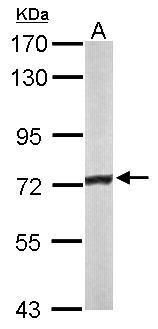
Sample (50 μg of whole cell lysate) A: mouse liver 7.5% SDS PAGE GTX114232 diluted at 1:3000 The HRP-conjugated anti-rabbit IgG antibody (GTX213110-01) was used to detect the primary antibody. |
|
|
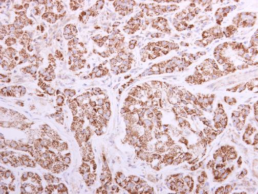
ACADVL antibody [N1C1] detects ACADVL protein at mitochondria by immunohistochemical analysis. Sample: Paraffin-embedded human A549 xenograft. ACADVL antibody [N1C1] (GTX114232) diluted at 1:500. Antigen Retrieval: Trilogy™ (EDTA based, pH 8.0) buffer, 15min |
|
|
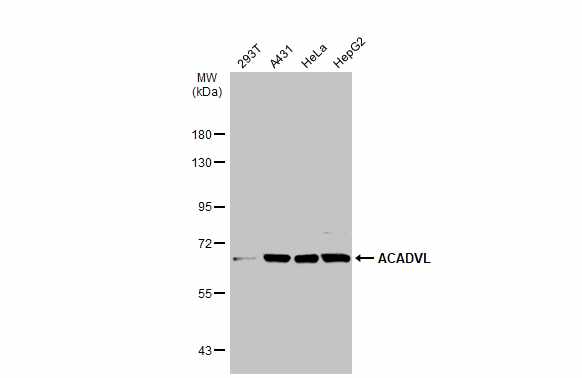
Various whole cell extracts (30 μg) were separated by 7.5% SDS-PAGE, and the membrane was blotted with ACADVL antibody [N1C1] (GTX114232) diluted at 1:1000. The HRP-conjugated anti-rabbit IgG antibody (GTX213110-01) was used to detect the primary antibody. |
|
|
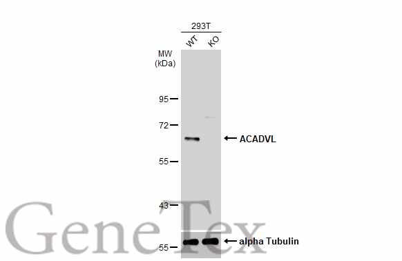
Wild-type (WT) and ACADVL knockout (KO) 293T cell extracts (30 μg) were separated by 7.5% SDS-PAGE, and the membrane was blotted with ACADVL antibody [N1C1] (GTX114232) diluted at 1:1000. The HRP-conjugated anti-rabbit IgG antibody (GTX213110-01) was used to detect the primary antibody. |
|
|
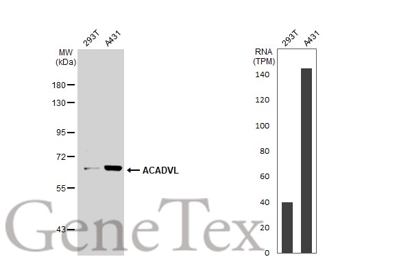
Various whole cell extracts (30 μg) were separated by 7.5% SDS-PAGE, and the membrane was blotted with ACADVL antibody [N1C1] (GTX114232) diluted at 1:1000. The HRP-conjugated anti-rabbit IgG antibody (GTX213110-01) was used to detect the primary antibody. Corresponding RNA expression data for the same cell lines are based on Human Protein Atlas program. |
|
|
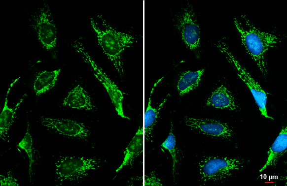
ACADVL antibody [N1C1] detects ACADVL protein at mitochondria by immunofluorescent analysis.Sample: HeLa cells were fixed in ice-cold MeOH for 5 min.Green: ACADVL stained by ACADVL antibody [N1C1] (GTX114232) diluted at 1:500.Blue: Fluoroshield with DAPI (GTX30920).Scale bar= 10 μm. |
|
|
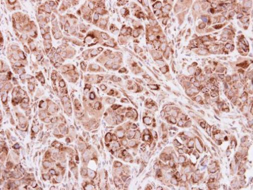
ACADVL antibody [N1C1] detects ACADVL protein at cytoplasm by immunohistochemical analysis.Sample: Paraffin-embedded mouse kidney.ACADVL stained by ACADVL antibody [N1C1] (GTX114232) diluted at 1:500.Antigen Retrieval: Citrate buffer, pH 6.0, 15 min |
|
|
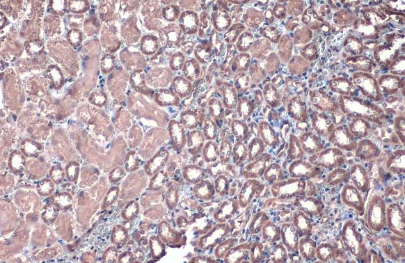
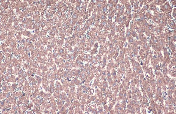
ACADVL antibody [N1C1] detects ACADVL protein at cytoplasm by immunohistochemical analysis.Sample: Paraffin-embedded rat liver.ACADVL stained by ACADVL antibody [N1C1] (GTX114232) diluted at 1:500.Antigen Retrieval: Citrate buffer, pH 6.0, 15 min |
Product Guarantee and Expert Support
