
GNAO1 antibody, Unconjugated, Rabbit, Polyclonal
Catalog Number:
GTX114439
- Images (9)
| Article Name: | GNAO1 antibody, Unconjugated, Rabbit, Polyclonal |
| Biozol Catalog Number: | GTX114439 |
| Supplier Catalog Number: | GTX114439 |
| Alternative Catalog Number: | GTX114439-100,GTX114439-25 |
| Manufacturer: | GeneTex |
| Host: | Rabbit |
| Category: | Antikörper |
| Application: | ICC, IHC-Fr, IHC-P, IP, WB |
| Species Reactivity: | Human, Mouse, Rat, Zebrafish |
| Immunogen: | Recombinant protein encompassing a sequence within the center region of human GNAO1. The exact sequence is proprietary. |
| Conjugation: | Unconjugated |
| Alternative Names: | G protein subunit alpha o1 , EIEE17 , G-ALPHA-o , GNAO , HLA-DQB1 , NEDIM |
| Application Notes: | WB: 1:500-1:20000. ICC/IF: 1:100-1:1000. IHC-P: 1:100-1:1000. IHC-Fr: 1:100-1:1000. IP: 1:100-1:500. *Optimal dilutions/concentrations should be determined by the researcher.Not tested in other applications. |
|
|
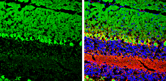
GTX114439 IHC-Fr Image |
|
|
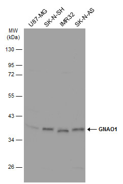
Various whole cell extracts (30 μg) were separated by 10% SDS-PAGE, and the membrane was blotted with GNAO1 antibody (GTX114439) diluted at 1:1000. The HRP-conjugated anti-rabbit IgG antibody (GTX213110-01) was used to detect the primary antibody. |
|
|
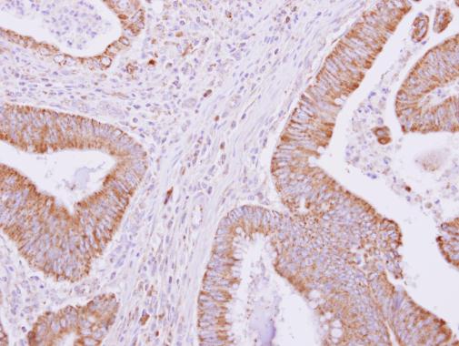
GNAO1 antibody detects GNAO1 protein at cytoplasm on human colon carcinoma by immunohistochemical analysis. Sample: Paraffin-embedded colon carcinoma. GNAO1 antibody (GTX114439) dilution: 1:500. Antigen Retrieval: Trilogy™ (EDTA based, pH 8.0) buffer, 15min |
|
|
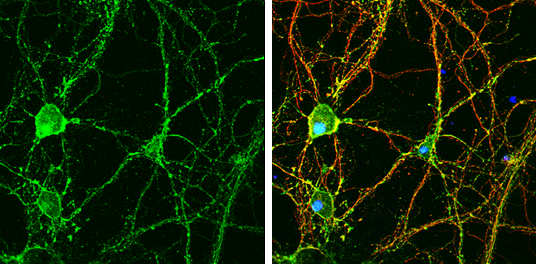
GNAO1 antibody detects GNAO1 protein by immunofluorescent analysis.Sample: DIV9 rat E18 primary hippocampal neuron cells were fixed in 4% paraformaldehyde at RT for 15 min.Green: GNAO1 stained by GNAO1 antibody (GTX114439) diluted at 1:500.Red: beta Tubulin 3/ Tuj1, stained by beta Tubulin 3/ Tuj1 antibody [GT1338] (GTX631831) diluted at 1:500.Blue: Fluoroshield with DAPI (GTX30920). |
|
|
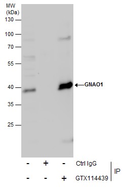
Immunoprecipitation of GNAO1 protein from HeLa whole cell extracts using 5 μg of GNAO1 antibody (GTX114439). Western blot analysis was performed using GNAO1 antibody (GTX114439). EasyBlot anti-Rabbit IgG (GTX221666-01) was used as a secondary reagent. |
|
|
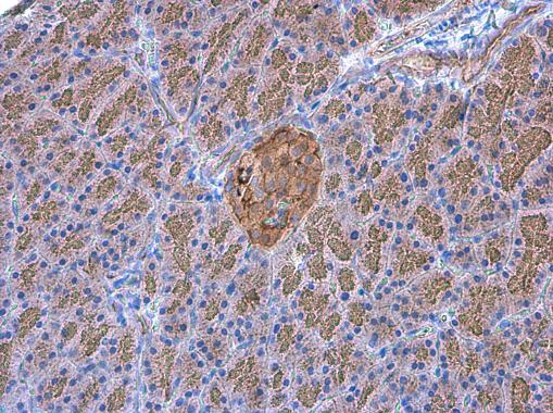
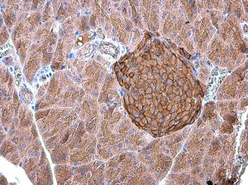
GNAO1 antibody detects GNAO1 protein at cell membrane and cytoplasm in rat pancreas by immunohistochemical analysis. Sample: Paraffin-embedded rat pancreas. GNAO1 antibody (GTX114439) diluted at 1:500. Antigen Retrieval: Citrate buffer, pH 6.0, 15 min |
|
|
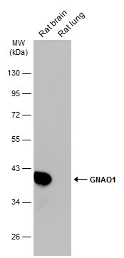
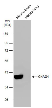
Various tissue extracts (50 μg) were separated by 10% SDS-PAGE, and the membrane was blotted with GNAO1 antibody (GTX114439) diluted at 1:10000. The HRP-conjugated anti-rabbit IgG antibody (GTX213110-01) was used to detect the primary antibody. |
|
|
GNAO1 antibody detects GNAO1 protein at cell membrane and cytoplasm in mouse pancreas by immunohistochemical analysis. Sample: Paraffin-embedded mouse pancreas. GNAO1 antibody (GTX114439) diluted at 1:500. Antigen Retrieval: Citrate buffer, pH 6.0, 15 min |
|
|
Various tissue extracts (50 μg) were separated by 10% SDS-PAGE, and the membrane was blotted with GNAO1 antibody (GTX114439) diluted at 1:10000. The HRP-conjugated anti-rabbit IgG antibody (GTX213110-01) was used to detect the primary antibody. |
Product Guarantee and Expert Support
