
Histone H1.0 antibody, Unconjugated, Rabbit, Polyclonal
Catalog Number:
GTX114462
- Images (9)
| Article Name: | Histone H1.0 antibody, Unconjugated, Rabbit, Polyclonal |
| Biozol Catalog Number: | GTX114462 |
| Supplier Catalog Number: | GTX114462 |
| Alternative Catalog Number: | GTX114462-100,GTX114462-25 |
| Manufacturer: | GeneTex |
| Host: | Rabbit |
| Category: | Antikörper |
| Application: | ICC, IHC-P, WB |
| Species Reactivity: | Human, Mouse, Rat |
| Immunogen: | Recombinant protein encompassing a sequence within the center region of human Histone H1.0. The exact sequence is proprietary. |
| Conjugation: | Unconjugated |
| Alternative Names: | H1 histone family member 0 , H10 , H1FV |
| Application Notes: | WB: 1:1000-1:20000. ICC/IF: 1:100-1:1000. IHC-P: 1:100-1:1000. *Optimal dilutions/concentrations should be determined by the researcher.Not tested in other applications. |
|
|
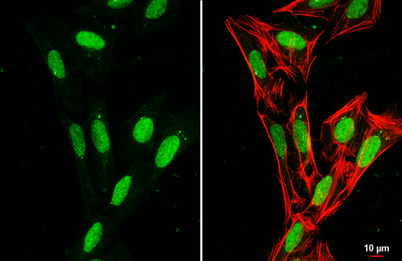
GTX114462 ICC/IF Image |
|
|
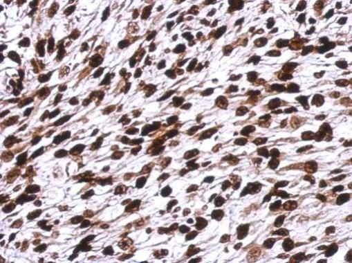
Immunohistochemical analysis of paraffin-embedded C2C12 xenograft, using Histone H1.0(GTX114462) antibody at 1:500 dilution. Antigen Retrieval: Trilogy™ (EDTA based, pH 8.0) buffer, 15min |
|
|
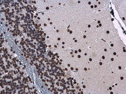
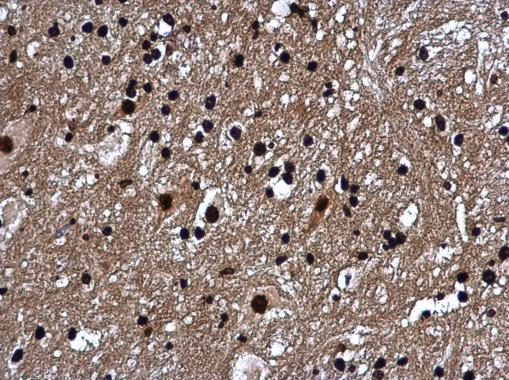
Histone H1.0 antibody detects Histone H1.0 protein at nucleus in mouse brain by immunohistochemical analysis. Sample: Paraffin-embedded mouse brain. Histone H1.0 antibody (GTX114462) diluted at 1:500. Antigen Retrieval: Citrate buffer, pH 6.0, 15 min |
|
|
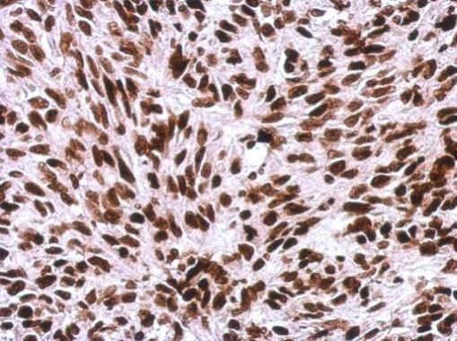
Immunohistochemical analysis of paraffin-embedded SkHep1 xenograft, using Histone H1.0(GTX114462) antibody at 1:500 dilution. Antigen Retrieval: Trilogy™ (EDTA based, pH 8.0) buffer, 15min |
|
|
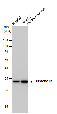
Histone H1 antibody detects Histone H1 protein by western blot analysis. HepG2 whole cell extracts and nuclear extracts (30 μg) were separated by 12% SDS-PAGE, and the membrane was blotted with Histone H1 antibody (GTX114462) at a dilution of 1:10000 and developed with Trident femto Western HRP Substrate (GTX14698). The HRP-conjugated anti-rabbit IgG antibody (GTX213110-01) was used to detect the primary antibody. |
|
|
Histone H1.0 antibody detects Histone H1.0 protein at nucleus in mouse brain by immunohistochemical analysis. Sample: Paraffin-embedded mouse brain. Histone H1.0 antibody (GTX114462) diluted at 1:500. Antigen Retrieval: Citrate buffer, pH 6.0, 15 min |
|
|
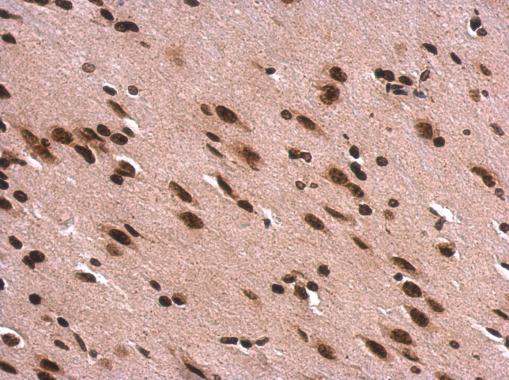
Histone H1.0 antibody detects Histone H1.0 protein at nucleus on rat fore brain by immunohistochemical analysis. Sample: Paraffin-embedded rat fore brain. Histone H1.0 antibody (GTX114462) dilution: 1:500. Antigen Retrieval: Trilogy™ (EDTA based, pH 8.0) buffer, 15min |
|
|
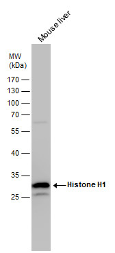
Histone H1 antibody detects Histone H1 protein by western blot analysis. Mouse tissue extracts (50 μg) was separated by 12% SDS-PAGE, and the membrane was blotted with Histone H1 antibody (GTX114462) diluted by 1:5000. The HRP-conjugated anti-rabbit IgG antibody (GTX213110-01) was used to detect the primary antibody. |
|
|
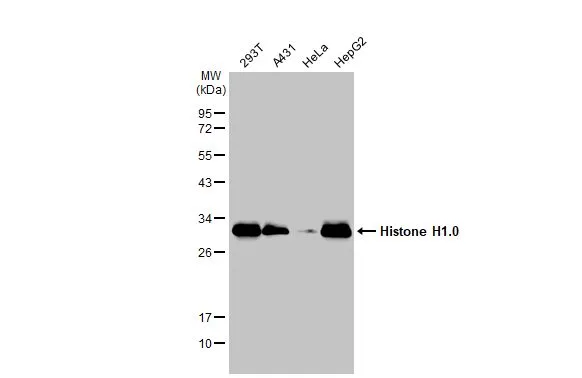
Various whole cell extracts (30 μg) were separated by 12% SDS-PAGE, and the membrane was blotted with Histone H1.0 antibody (GTX114462) diluted at 1:10000. The HRP-conjugated anti-rabbit IgG antibody (GTX213110-01) was used to detect the primary antibody. |
Product Guarantee and Expert Support
