Perforin is a pore-forming protein that leads to osmotic lysis of the target cells and subsequently enables granzymes to enter the target cells and activate apoptosis. Perforin has structural and functional similarities to complement component 9 (C9). Like C9, this protein creates transmembrane tubules and is capable of lysing non-specifically a variety of target cells. It is one of the main cytolytic proteins of cytolytic granules, and is known to be a key effector molecule for T-cell- and natural killer-cell-mediated cytolysis. Defects in this gene cause familial hemophagocytic lymphohistiocytosis type 2 (HPLH2), a rare and lethal autosomal recessive disorder of early childhood. The expression of perforin is reportedly upregulated in activated CD8+ T-cells, natural killer cells and some CD4+ T-cells.
200ug/ml of Ab Purified from Bioreactor Concentrate by Protein A/G. Prepared in 10mM PBS with 0.05% BSA & 0.05% azide. Also available WITHOUT BSA & azide at 1.0mg/ml.
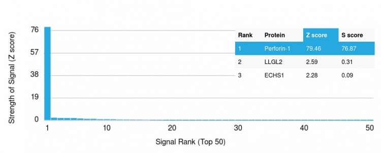
Analysis of Protein Array containing more than 19,000 full-length human proteins using Perforin-1 Mouse Monoclonal Antibody (PRF1/2468) Z- and S- Score: The Z-score represents the strength of a signal that a monoclonal antibody (Monoclonal Antibody) (in combination with a fluorescently-tagged anti-IgG secondary antibody) produces when binding to a particular protein on the HuProtTM array. Z-scores are described in units of standard deviations (SDs) above the mean value of all signals generated on that array. If targets on HuProtTM are arranged in descending order of the Z-score, the S-score is the difference (also in units of SDs) between the Z-score. S-score therefore represents the relative target specificity of a Monoclonal Antibody to its intended target. A Monoclonal Antibody is considered to specific to its intended target, if the Monoclonal Antibody has an S-score of at least 2.5. For example, if a Monoclonal Antibody binds to protein X with a Z-score of 43 and to protein Y with a Z-score of 14, then the S-score for the binding of that Monoclonal Antibody to protein X is equal to 29.
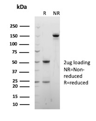
SDS-PAGE Analysis of Purified Perforin-1 Mouse Monoclonal Antibody (PRF1/2468). Confirmation of Purity and Integrity of Antibody.
SDS-PAGE Analysis of Purified Perforin-1 Recombinant Mouse Monoclonal Antibody (PRF1/2468). Confirmation of Purity and Integrity of Antibody.
* VAT and and shipping costs not included. Errors and price changes excepted