Protein Kinase C iota / lambda / PRKCI Antibody, IgG2b, Clone: [PRKCI/4912], Mouse, Monoclonal
Biozol Catalog Number:
NBT-5584-MSM2-P1ABX
Supplier Catalog Number:
5584-MSM2-P1ABX
Alternative Catalog Number:
NBT-5584-MSM2-P1ABX-100
Manufacturer:
NeoBiotechnologies
Host:
Mouse
Category:
Antikörper
Application:
IHC
Species Reactivity:
Human
Immunogen:
Recombinant fragment (around aa100-300) of human PRKCI protein (exact sequence is proprietary)
Alternative Names:
aPKC lambda/iota, Atypical protein kinase C lambda/iota, Atypical protein kinase C-lambda/iota, nPKC-iota, nPKCiota, PKC lambda/iota, PKCI, PKCiota, PKClambda, PRKC iota, PRKC lambda/iota, PRKCI, Protein kinase C iota type
Members of the protein kinase C (PKC) family play a key regulatory role in a variety of cellular functions, including cell growth and differentiation, gene expression, hormone secretion and membrane function. PKCs were originally identified as serine/threonine protein kinases whose activity was dependent on calcium and phospholipids. Diacylglycerols (DAG) and tumor promoting phorbol esters bind to and activate PKC. PKCs can be subdivided into at least two major classes, including conventional (c) PKC isoforms ((TM), A°aI, A°aII and A) and novel (n) PKC isoforms ( OOL, , OOEÖ, , , l/i, m and n). Patterns of expression for each PKC isoform differ among tissues and PKC family members exhibit clear differences in their cofactor dependencies. For instance, the kinase activities of PKC OOL and are independent of Ca2+. On the other hand, most of the other PKC members possess phorbol ester-binding activities and kinase activities.
200ug/ml of Ab purified from Bioreactor Concentrate by Protein A/G. Prepared in 10mM PBS with 0.05% BSA & 0.05% azide. Also available WITHOUT BSA & azide at 1.0mg/ml.
Antibody Type:
Monoclonal Antibody
Application Notes:
Immunohistochemistry (Formalin-fixed) (1-2ug/ml for 30 minutes at RT),(Staining of formalin-fixed tissues requires heating tissue sections in 10mM Tris with 1mM EDTA, pH 9.0, for 45 min at 95 °C followed by cooling at RT for 20 minutes),Optimal diluti
Formalin-fixed, paraffin-embedded human brain stained with Protein Kinase C iota / lambda Mouse Monoclonal Antibody (PRKCI/4912).
SDS-PAGE Analysis of Purified PRKCI Mouse Monoclonal Antibody (PRKCI/4912). Confirmation of Purity and Integrity of Antibody.
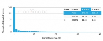
Analysis of Protein Array containing more than 19,000 full-length human proteins using Protein Kinase C iota / lambda Mouse Monoclonal Antibody (PRKCI/4912). Z- and S- Score: The Z-score represents the strength of a signal that a monoclonal antibody (MAb) (in combination with a fluorescently-tagged anti-IgG secondary antibody) produces when binding to a particular protein on the HuProtTM array. Z-scores are described in units of standard deviations (SDs) above the mean value of all signals generated on that array. If targets on HuProtTM are arranged in descending order of the Z-score, the S-score is the difference (also in units of SDs) between the Z-score. S-score therefore represents the relative target specificity of a MAb to its intended target. A MAb is considered to specific to its intended target, if the MAb has an S-score of at least 2.5. For example, if a MAb binds to protein X with a Z-score of 43 and to protein Y with a Z-score of 14, then the S-score for the binding of that MAb to protein X is equal to 29.
* VAT and and shipping costs not included. Errors and price changes excepted