Recombinant fragment (around aa29-148) of human RBP4 protein (exact sequence is proprietary)
Alternative Names:
Plasma retinol binding protein 4 (PRBP), RDCCAS, Retinol binding protein 4 interstitial
Retinol (Vitamin A) is transported in the blood bound to its carrier protein, retinol-binding protein (RBP), also designated plasma retinol-binding protein (PRBP) or RBP4. A member of the lipocalin family, RBP conveys retinol from stores in the liver to peripheral tissues. In plasma, RBP binds transthyretin (TTR, formerly called prealbumin) to prevent glomerular filtration of low molecular weight RBP in the kidneys. The stability of this complex holds diagnostic importance because the molar ratio of RBP:TTR provides an indirect way to indicate marginal Vitamin A deficiency. Vitamin A deficiency blocks the secretion of RBP, resulting in defective delivery and supply to epidermal cells. Originally identified solely as a transporter protein, recent studies correlating increased levels of RBP expression in adipose tissue with Insulin resistance have generated research into the possible roles the protein may play in the pathogenesis of type 2 diabetes and obesity.
200ug/ml of Ab purified from Bioreactor Concentrate by Protein A/G. Prepared in 10mM PBS with 0.05% BSA & 0.05% azide. Also available WITHOUT BSA & azide at 1.0mg/ml.
Antibody Type:
Monoclonal Antibody
Application Notes:
Immunohistochemistry (Formalin-fixed) (1-2ug/ml for 30 minutes at RT),(Staining of formalin-fixed tissues requires heating tissue sections in 10mM Tris with 1mM EDTA, pH 9.0, for 45 min at 95 °C followed by cooling at RT for 20 minutes),Optimal diluti
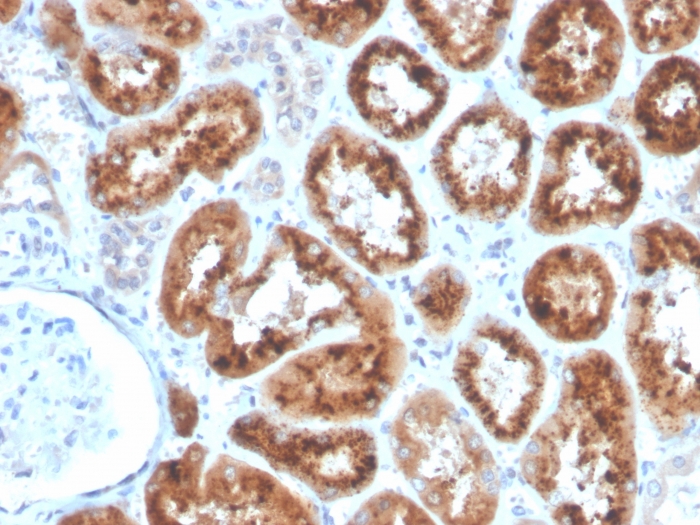
Formalin-fixed, paraffin-embedded human kidney stained with RBP4 Mouse Monoclonal Antibody (RBP4/4053).
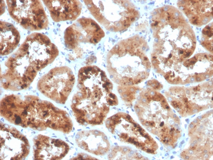
Formalin-fixed, paraffin-embedded human kidney stained with RBP4 Mouse Monoclonal Antibody (RBP4/4053).
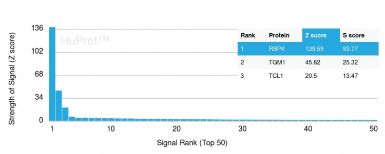
Analysis of Protein Array containing more than 19,000 full-length human proteins using RBP4 Mouse Monoclonal Antibody (RBP4/4053). Z- and S- Score: The Z-score represents the strength of a signal that a monoclonal antibody (MAb) (in combination with a fluorescently-tagged anti-IgG secondary antibody) produces when binding to a particular protein on the HuProtTM array. Z-scores are described in units of standard deviations (SDs) above the mean value of all signals generated on that array. If targets on HuProtTM are arranged in descending order of the Z-score, the S-score is the difference (also in units of SDs) between the Z-score. S-score therefore represents the relative target specificity of a MAb to its intended target. A MAb is considered to specific to its intended target, if the MAb has an S-score of at least 2.5. For example, if a MAb binds to protein X with a Z-score of 43 and to protein Y with a Z-score of 14, then the S-score for the binding of that MAb to protein X is equal to 29.
Western Blot Analysis of human kidney tissue lysate using RBP4 Mouse Monoclonal Antibody (RBP4/4053).
* VAT and and shipping costs not included. Errors and price changes excepted